
|
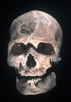
I had the opportunity contribute to two articles for a special issue of Quaternary International entitled "Not Only Use" that was edited by Juan Luis Fernández-Marchena, Lena Asryan, Antonella Pedergnana, and Andreu Ollé. The issue contains an impressive overview various methodologies used to study wear the burgeoning interest in multidisciplinary efforts to study wear traces (i.e., traceology) of different origins and the processes (i.e., operative chains) involved in manufacture, use, and abandonment of an object that is later recovered by an archaeologist (check out the editorial by Fernández-Marchena et al. for more info). The first article was lead by Raquel Hernando. In short, Hernando was interested in understanding if recent advances in optical microscopy could resurrect its use for the study of human dental microwear. Hernando noted that dental microwear analyses originally used optical microscopy, but it was later replaced by scanning electron microscopy (SEM). Eventually confocal microscopy became the favored technology for occlusal dental microwear analysis (i.e., dental microwear texture analysis, or "DMTA"), while SEM is still preferred for buccal microwear analysis. However, Hernando and colleagues note that SEM analyses are costly (we generally pay by the hour to use these microscopes) and the postprocessing of images is also quite time consuming (and exhausting for your eyes!). DMTA is generally quicker, but the microscopes—and software needed for DMTA—is not nearly as widely accessible as SEM. That means costly travel, lodging, and user fees to do DMTA analyses for many of us without local access to equipment. So, why not revisit optical microscopy? Hernando and colleagues point to many advances in optical microscopy that have been explored in the context of traceology. Buccal microwear seemed like the best place to start since it still widely uses SEM. Hernando and colleagues found that OM produces very similar results to traditional SEM methods whether one studies the original tooth or a dental cast of a tooth (see image below). 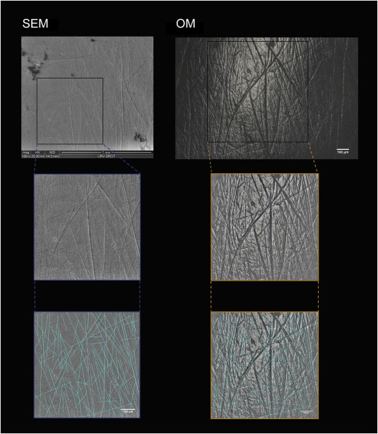 Above: Comparison of scanning electron (SEM) and optical microscopy (OM) images of buccal microwear. Note the excellent resolution in OM. Raquel Hernando and colleagues noted that optical microscopy provides many other advantages over traditional SEM analysis: less expensive equipment with less associated maintenance, wider accessibility of optical microscopes for researchers, less eye fatigue, greater image resolution, 3D appearance of images with greater definition, and relatively quick data acquisition and analysis. A drawback is the need to build up open-access databases for comparitive purposes, but the data produced in this paper marks the beginning of that effort. This study points out the importance of revisiting methodologies with a critical eye, but also how interdisciplinary research—something IPHES takes great pride in—can lead to innovation in allied fields of research. The second article explored the use of gigapixel-like (GPL) images for studying external surfaces of teeth. GPL images make use focus-stacking (extended focus images) and panoramic stitching of microscopic images to create mosaic images with high depth of field using SEM. This “gigapixel-like” (GPL) imaging strategy can be used to create multiscale, high-resolution images of entire, or partial, dental surfaces that can be viewed from a field of view that encompasses an entire tooth surface to high magnification views of dental microstructure, microwear, taphonomic features, among other features. The images have a variety of uses from the communication of results in scientific publications to their use in interactice museum displays and websites or training researchers. 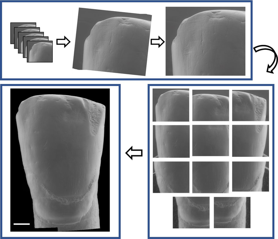 Above: simplified outline of focus-stacking and creation of image mosaic to create a gigapixel-like (GPL) image. 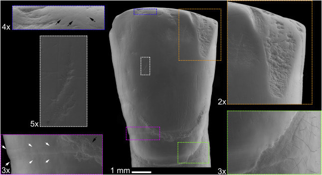 Above: A GPL image (center) with call-out boxes of varying magnification that indicate different surface features. Descriptions proceed clockwise from upper right corner. Orange rectangle: Medium size antemortem enamel chip with well-worn margins. Green rectangle: Detail of cementoenamel junction and root surface. Subtle perikymata (bottom left quadrant) and striations (upper left quadrant) are visible on the enamel. Subtle postmortem cracking of root surface also evident. Magenta rectangle: Detail of furrow-form hypoplasia with clearly visible perikymata (between white arrows). Black arrow points to dental calculus deposit. White rectangle: Detail of instrumental striation with a right oblique orientation. Blue rectangle: arrows indicate microstriations on labioincisal edge and a well-worn, but small, antemortem enamel chip to the left of the image. While the goal of the publication was to outline the GPL methodology and uses, we also made an interesting discovery from the creation of a GPL image for one of the teeth from the Chalcolithic context (dated to about 4000 years before present) of El Mirador Cave near Burgos, Spain. We found that at least one tooth exhibited a strange discoloration when viewed with the naked eye (see photo below). Microscopic examination revealed that the discoloration is related to enamel erosion—something that is rarely documented in prehistoric contexts. 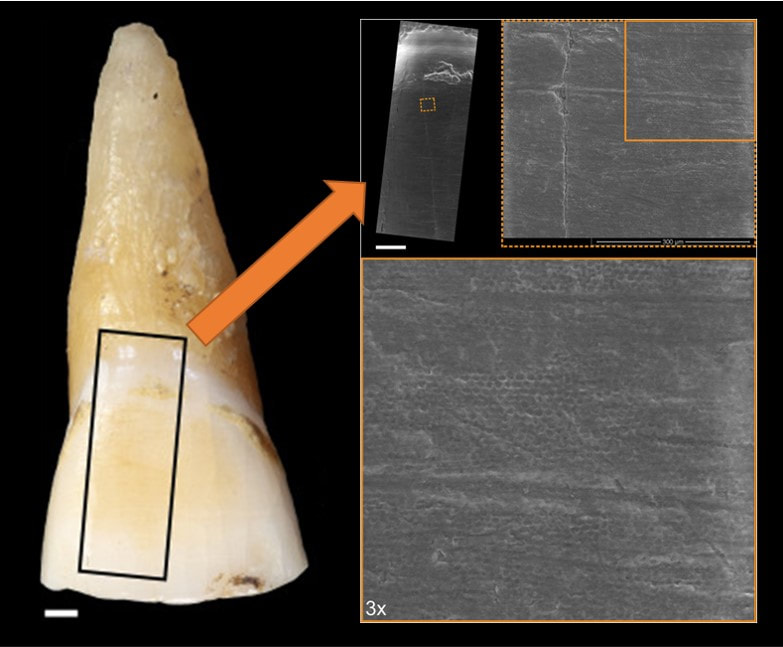 Above: Photo of original tooth with discolored (yellowish) enamel surface. GPL image sampling indicated by black box and GPL image indicated by orange arrow. Zooming in on section 300x shows "honey-comb" appearance of enamel surface. This indicates erosion of the enamel. This study makes me suspect that erosion in teeth from archaeological contexts is much higher than we currently acknowledge, and calls for a need for detailed analyses of the original teeth in conjunction with high magnification analysis for definitive diagnosis. Nonetheless, this is a very interesting (and rather accidental) discovery. More analyses of the El Mirador material are underway. References and further reading These studies:
Additional references:
0 Comments
There is a deep history of archaeological investigation focusing on the Bronze Age El Argar, or Argaric, cultural phenomenon from southeastern Spain. Argaric archaeology is probably most famous for the elaborate settlement structures, well-preserved burials, and evidence for sophisticated metallurgy and material culture. The rich archaeological record and excellent preservation of human remains have provided archaeologists with incredible resources for reconstructing the lifeways of these Bronze Age peoples.
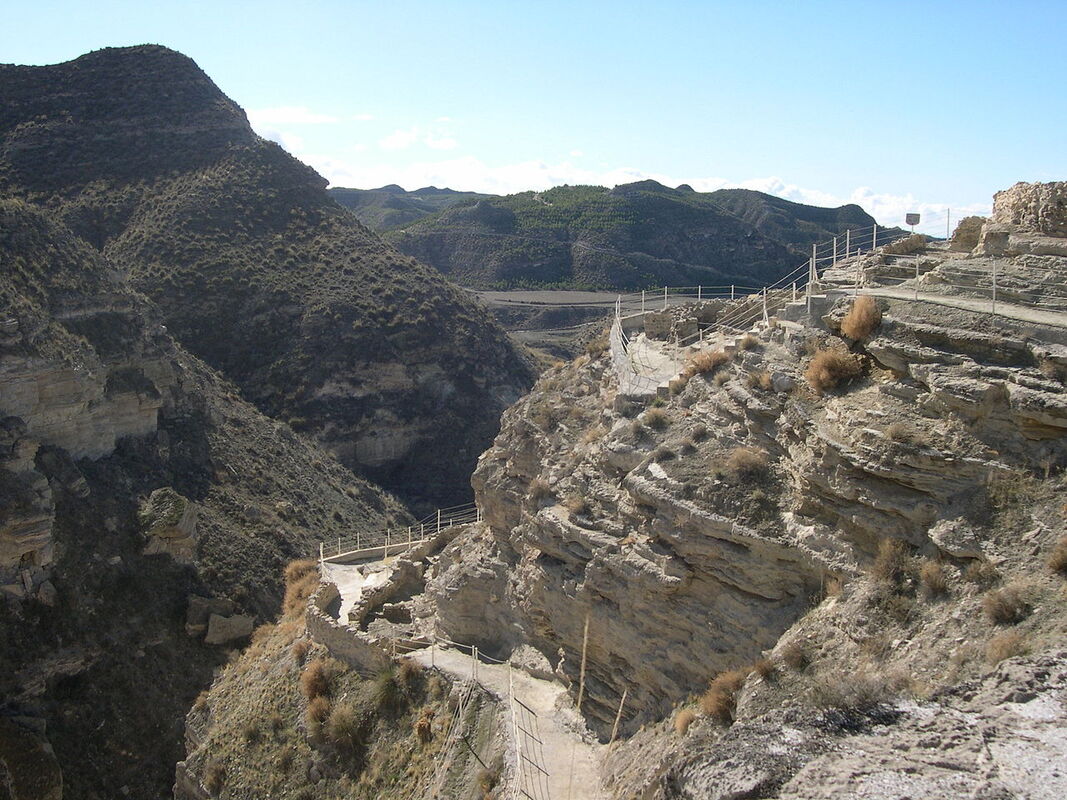
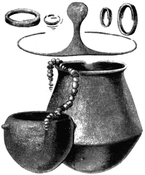
Above Left: view of the site of Castellón Alto. By Rafael Jiménez from Córdoba, España - Castellon Alto 2, CC BY-SA 2.0, https://commons.wikimedia.org/w/index.php?curid=21664792. Above Right: examples of grave goods recovered from an Argaric context. By Luis Siret - Siret, H., and Louis Siret (1887). «Les premiers âges du métal dans le sud-est de l'Espagne». Anvers., Public Domain, https://commons.wikimedia.org/w/index.php?curid=1294319 I was recently involved in a collaborative research project on the human remains from the site of Castellón Alto that focused on the dental remains of the individuals buried at the site. As part of his Ph.D. research on the human remains from the site, Àngel Rubio discovered an interesting trend at the site: of the 106 burials examined, the teeth of 5 individuals showed atypical patterns of dental wear (see below). What was even more astounding was that each of those individuals was female. No males had these interesting patterns of wear. Further microscopic analysis conducted by Dr. Marina Lozano provided clues as to what behaviors may have contributed to the unique wear patterns identified on the teeth of these 5 individuals. 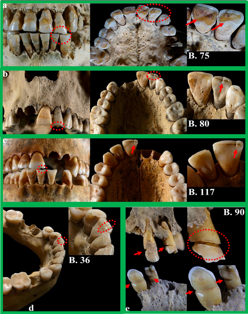 Above: The 5 female individuals with atypical patterns of dental wear. Red arrows and circles indicate the location of the wear in the photos. Image from the article: https://doi.org/10.1016/j.jas.2020.105239. The remarkable preservation of organic remains (textiles, wool, plant fibers, etc.) in addition to extensive durable material culture (awls, loom weights, spindle whorl, needles) found at Argaric archaeological sites provided additional clues as to what tasks may have contributed to the unique patterns of dental wear on the 5 women from Castellón Alto. A probable explanation is that at least some of the women at the site were involved in specialized craft production such as textile production, processing of fiber or cordage, basketry, and similar tasks (see illustration below). Ethnohistoric documentation of the use of the teeth for craft production adds additional support that the formation of atypical dental wear in the subset of the women from Castellón Alto was related to craft production. 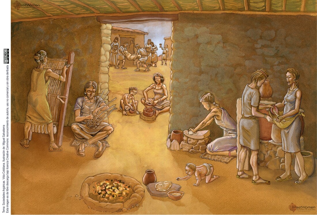 Above: A scene of Argaric life featuring the many of the tasks related to food preparation and craft-production in the foreground. Ilustración: Miguel Salvatierra "Cultura argárica". The rich archaeological record from the Argaric contexts of southeasten Spain is bound to reveal more insights into human social lives and identities of Bronze Age peoples. In this case, analyses have revealed a unique role for at least some of the women buried at the site engaged in. References and further readingThis study:
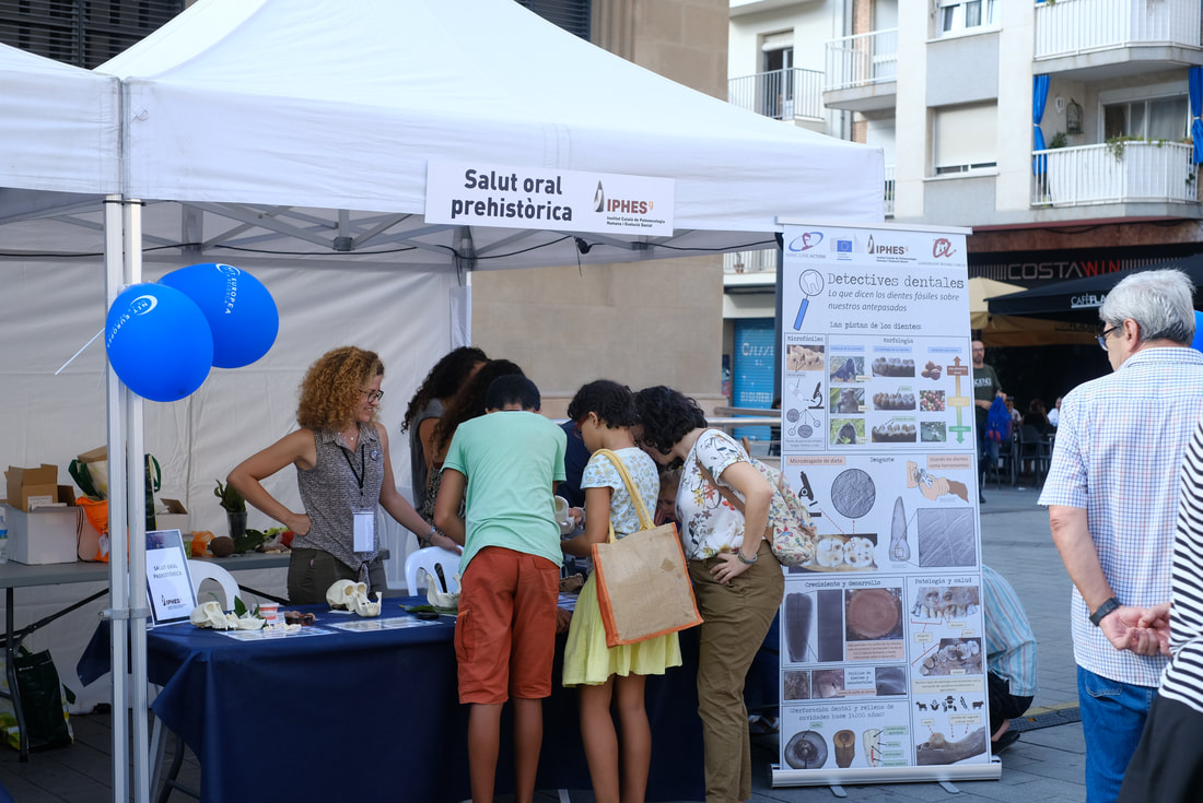
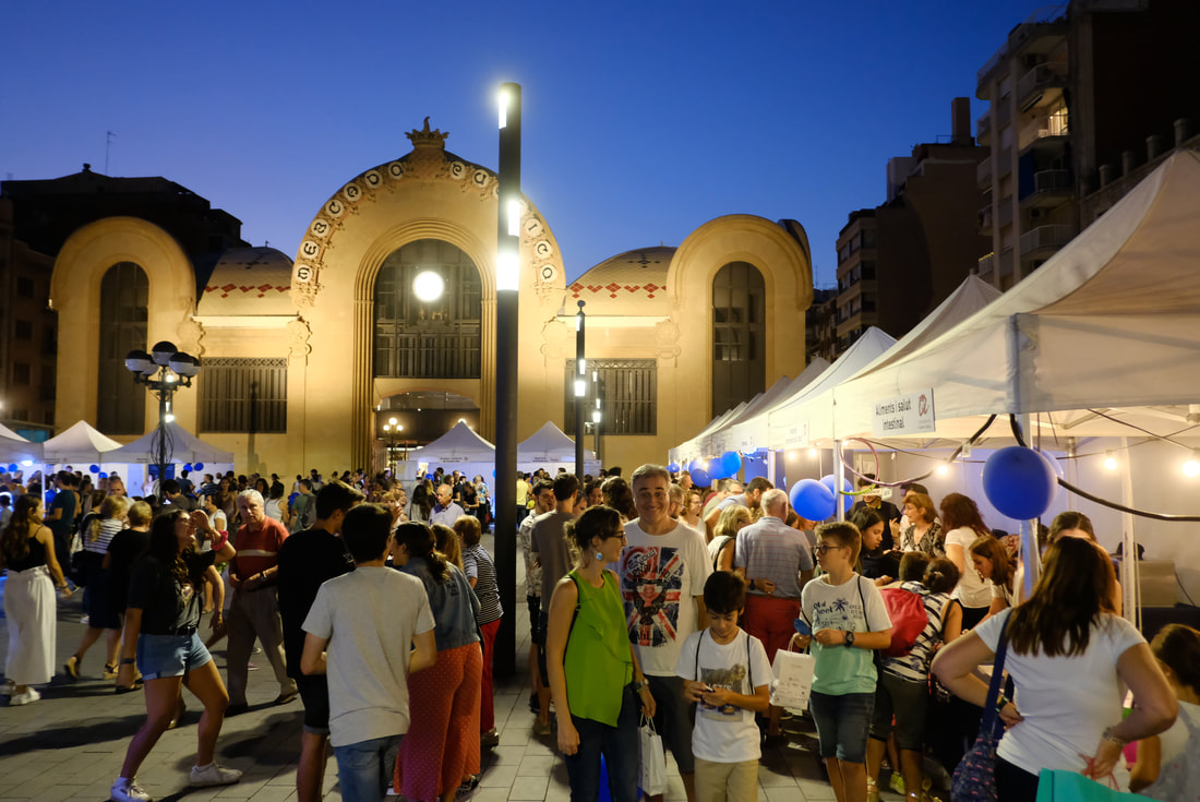
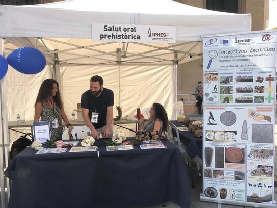
Lozano M, Jiménez-Brobeil SA, Willman JC, Sánchez-Barba LP, Molina F, and Rubio Á. 2020. Argaric craftswomen: Sex-based division of labor in the Bronze Age southeastern Iberia. Journal of Archaeological Science:105239. https://doi.org/10.1016/j.jas.2020.105239. Additional references: I must emphasize the great resources at http://www.pastwomen.net/ and the resources on the Argaric culture in particular (http://www.pastwomen.net/objetos/cultura-argarica) for the preparation of this post. The website offers incredible, multi-language resources for the instructors, scholars, and anyone that is curious about the social lives of women in the past. The researchers, artists, and other contributors have really created an incredible resource. A little over two weeks ago was the Nit Europea de la Recerca (European Researcher’s Night) in Tarragona. It was part of a massive initiative that involved events in over 300 cities across Europe. The theme centered on International Year of the Periodic Table. Several of my IPHES colleagues and I (Institut Català de Paleoecologia Humana i Evolució Social, IPHES) participated in the event alongside numerous researchers from the Universitat Rovira i Virgili (URV), Catalan Institute of Classical Archaeology (Institut Català d’Arqueologia Clàssica, ICAC), the Pere Virgili Institute of Health Research (Institut d'Investigació Sanitària Pere Virgili, IISPV), and Institute of Chemical Research of Catalonia (Institut Català d'Investigació Química, ICIQ) provided exciting and unique scientific outreach activities for the children and families. The event was free and took place in front of the central market (Plaça Corsini) in Tarragona. Tables and booths were set up for all the research groups and the children started pouring in around 4:30.  Our contribution was Detectives Dentales: Lo que dicen los dientes fósiles sobre nuestros antepasados (Dental Detectives: What fossil teeth say about our ancestors). Like the rest of the researchers, we had a booth with tables set up, hands-on activities for the kids, and a giant poster describing our research. Hundreds of children came to our table to learn about human evolution, primates, hominins, and bioarchaeology over the course of the night. Thanks to the support and help of my incredible colleagues (Marina Lozano, Raquel Hernando, Efstathia Robakis, Miquel Guardiola Fígols, and Marta Fontanals Torroja), and the organizers of the event, everything went incredibly smoothly. ComCiència URV (@cienciaURV) also provided some great pictures of the event: Admittedly, I was terrified of doing this event. My Castellano is terrible, and my Catalan is non-existent, which is a constant source of stress and embarrassment for me. While my colleagues made sure there were no horrible mistakes in the materials we used (poster, fact sheets, etc.), there was still the issue of needing to speak Castellano for about 5 hours. Thankfully, I had incredible help with the event from my colleagues. And then there were the parents and the children… Simply put, they were incredible! I spoke in Castellano (as best I could) for most of the event, but many parents would extrapolate what I was communicating to their children or translate it into Catalan. In fact, there was a lot of translation. Examples I can immediately recall included translating my Castellano to Chinese and Italian. Other children that were not originally from Spain, were more comfortable with English than Castellano, so I obliged. Some of these children even translated what I said (in English) to their mother tongue for their younger siblings – examples included German, Italian, and Chinese. Some children would just look at me puzzled when I spoke, then took a look at my name tag (misspelled “John Williams” for the night, but still very much not a Spanish name), which prompted them to ask, “Can you say something in English?” I wasn’t prepared for this, but it was quite funny, and gave some children a chance to practice English. Another common end to an interaction was a “thank-you” in English from parents as they moved on to another table – a kind of wink and nod that they appreciated the effort. All in all, was surprised at how well everything went despite my initial anxieties concerning language barriers. Some reflections on this type of public outreach event I wanted to share a few reflections on what worked well for this type of outreach event, since many of my colleagues are involved in these sorts of activities. I envisioned setting this event up much like the “fossil laboratory” events that I used to set up for Introduction to Human Evolution / Biological Anthropology courses when I was a graduate student – albeit, this was for a much younger audience! The target age group was around 5-12 year olds but we definitely had some younger children too. And to be honest, many of the parents showed an interest in the materials on par with, and sometimes exceeding, that of their children! So here’s what worked and didn’t:
An enormous poster: We had a giant poster with lots of examples, or “clues”, that biological anthropologists use to understand and interpret primate and hominin dental morphology, diet, growth and development, behavior, pathology, etc. By “giant”, I mean 200 x 80 cm… About three times larger than any conference poster I’ve ever made… It was a monster. Keep in mind that Powerpoint will not make posters this big, so a half-size (100 x 40 cm) poster was made. I had to constantly remind myself to zoom in 200% to get an accurate sense of whether or not each image would be blurry or if the text was too small. In the end, the poster turned out really well and will be used for future science communication events at IPHES since it was printed on durable vinyl.
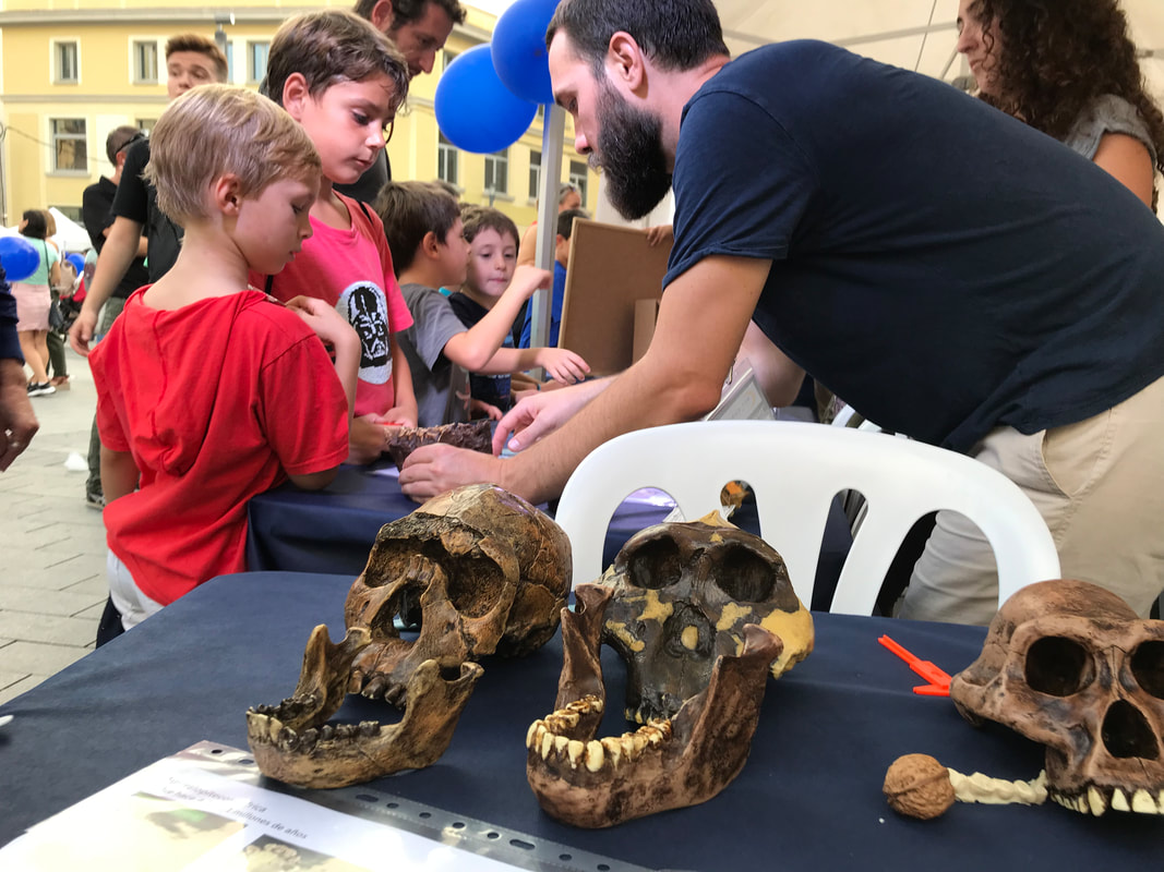
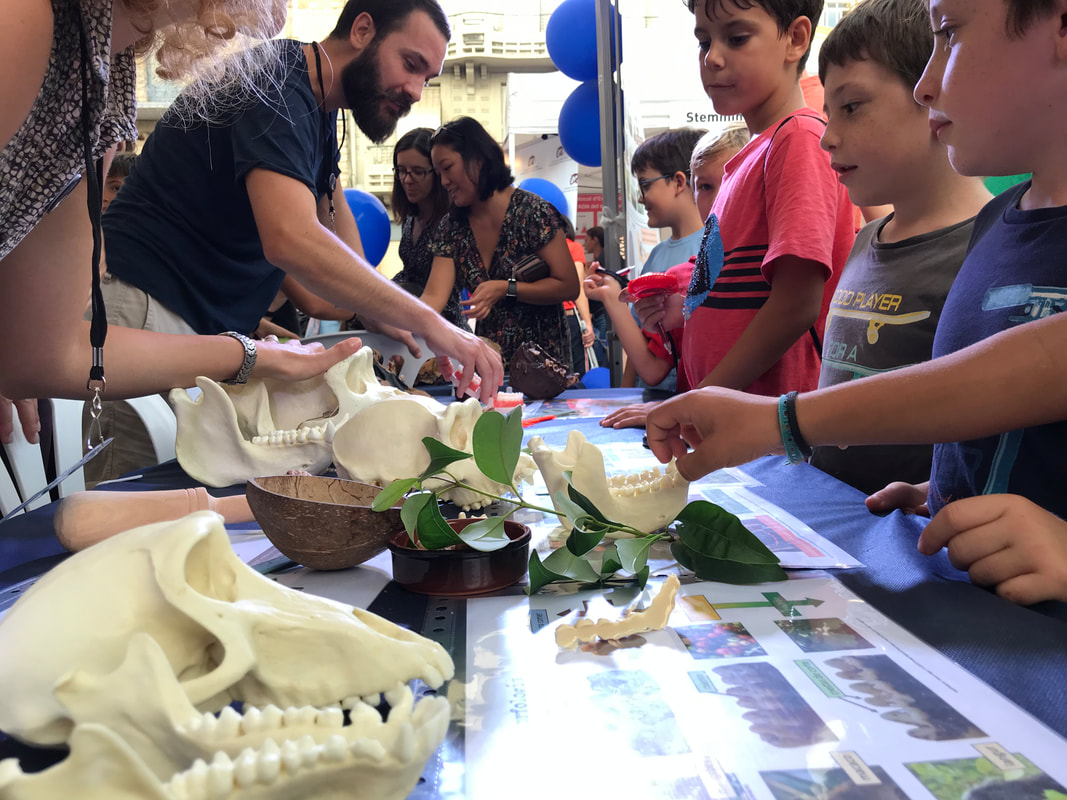
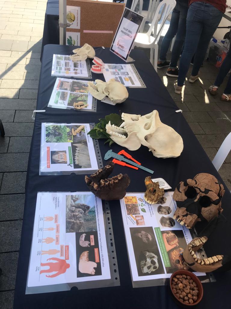
Primate and hominin casts (“No these are not the original fossils”): We set up a variety of casts from the URV/IPHES teaching collections to illustrate various aspects of primate functional morphology and hominin cranial and dental evolution. What I didn’t know would turn out to be so useful were the laminated sheets that accompanied each cast or groups of casts. For example, sheets for living primates included bright and colorful images of living primates eating and some basic descriptions of how their teeth help break down certain types of food. The hominin casts that we used included examples of Australopithecines, Paranthropus, Homo erectus, archaic Homo – a Neandertal and the Sima de los Huesos Skull 5 (“Miguelón”), and a recent modern human. Every cast also had an information sheet with a few facts about the fossil and reconstructions to help the kids visualize what these hominins may have looked like in life (The Smithsonian Institution, as always, was a great resource for this). The kids really liked the Gigantopithecus blacki mandible, but this was partly due to the comic relief provided by the body size comparisons in the information sheet we provided. The laminated sheets were not taped down, which also helped a lot. We were often completely surrounded by kids and unable to move around the tables with ease. Instead, being able to reach and grab the facts sheets and casts from across the table proved very helpful.
Food breakdown: To help demonstrate some of the ways that teeth breakdown food, we had some props on the table to help with explanations. We used dried figs as our fruit example since they are a much less messy alternative to fresh fruit. We paired the fruit with a mortar and pestle to discuss bunodont molars. We had plenty of leaves on the table, and we discussed shearing crests by miming the cutting action of scissors with our fingers. Originally, I was going to have real scissors to cut leaves but quickly realized how dangerous that might be as children started swarming the table! We also had walnuts (hard food), a nutcracker, and chufa (tough, high fiber food) on display. The chufa was one of my personal favorites and it was easily identified by parents (and some kids) as an ingredient for horchata. Aside from being delicious, chufa is a hypothetical C4 plant food of Paranthropus bosei – original article here). My colleague, Dr. Erin Kane, uses similar strategies in her course on the “Evolution of the Human Diet” which inspired me to go ahead with this idea at Researcher’s Night. She used a variety of other tools and foods for her demonstrations - like a staple remover to demonstrate how some teeth are well-suited for piercing the exoskeletons of insects - which are great for classroom settings. Like the scissors, we couldn't incorporate all the examples that Dr. Kane uses, but I can't wait to expand this activity the next time I get to teach this section in my own course. Dr. Kane also explores oral processing of food in her class with a variety of snacks – another thing I can’t wait to adapt for outreach events and teaching in the future.
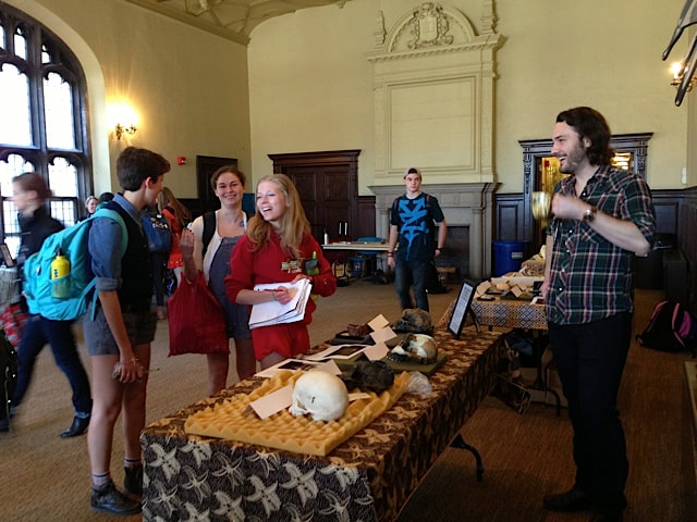
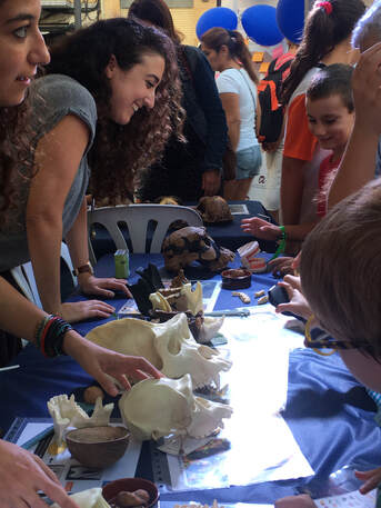
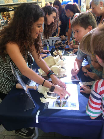
My colleagues, Raquel Hernando and Efstathia Robakis, teaching children about non-human primates and early hominins. If you look closely you will find a child playing with the loop. Other odds n’ ends: I molded the teeth of each fossil cast that we had on display and made dental casts in advance of the event out of a cheap and durable polyurethane resin. These casts were a lot easier for smaller children to handle. I also put a few 10x magnifying loops on the table and a handful of small plastic calipers. I never would have thought they’d be so popular, but many children measured everything they could – from the teeth and jaws on the table to the smile of a friend. The loops were even more popular and worked perfectly for examining the extra dental casts I made. One young scientist, I’m guessing she was 4 years old, came back to the table at least 3 times over the course of the evening just to use the calipers and loops! I rarely left my home without a loop when I was that young, and it is nice to see that these cheap little devices still fascinate children in an age of touchscreens.
What I would do differently: One of my big regrets centered around not having souvenirs for the children. This was largely due to a lack of time on my part and it’s something I definitely want to be better prepared for in the future. The original idea was to create a large sample of hemi-mandibles or hemi-maxillas out of dental stone. A variety of non-human primate and hominin teeth would be made, and each cast would come in a small bag with a fact card describing morphology, diet, etc. It’s not much, but I remember loving these sorts of things as a kid, and I would have liked to have had them ready for the kids at Researcher's Night. Alas, several hundred dental casts required quite a lot more time than I had to spare prior to Researcher’s Night. More loops. I ordered a 10 pack of loops but only one unit arrived, and I only had one other I was willing to risk damage to. The kids really liked them, so next time I will be better prepared with a dozen or so cheap loops spread across the tables. I’m sure I’ll continue to think of other things I’d wish to change but I think I’ll save that for the aftermath of the next public outreach event I take part in. All in all, this was one of the most unique and rewarding events during my postdoctoral experience in Tarragona. Again, this is in large part to the incredible help provided by my colleagues and the enthusiasm of the children and families that attended Researcher's Night. Many thanks to all! Last week I had the opportunity to attend the XVIIe Congrès Mondial UISPP - Union Internationale des Sciences Préhistoriques et Protohistoriques in Paris, France where I participated in Session XXXI-1 Through time, space and species: implication of new discoveries, technological developments and data diffusion improvement in Biological Anthropology organized by Dominique Grimaud-Hervé, Carlos Lorenzo, Julie Arnaud. I presented collaborative work synthsizing research on the "Non-alimentary tooth-use in European Prehistory". The presentation brought together data across a wide span of time and space that have been studied so far by my colleagues (Alejandro Romero, Eulàlia Subirà, and Marina Lozano) and me. Much of the data we presented will contribute to the IDENTITIES Project. 
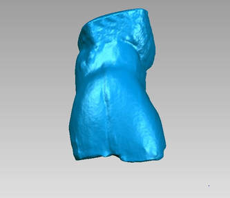
I am currently on a research trip at the Universidad de Alicante where I am working with Alejandro Romero on 3D methdologies for the IDENTITIES Project. The campus and weather are beautiful to say the least. 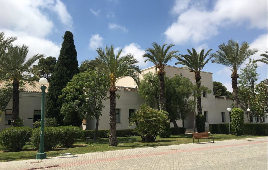 Departamento de Biotecnología, Universidad de Alicante where I am currently working. My task for the two weeks that I am in Alicante is to scan as many teeth as possible and begin to work with the software needed to manipulate and analyze the 3D models we create. Below you can see the scanner in the background and a dental cast sitting on a rotating platter. The platter rotates automatically and scans are taken at set increments. The scans are then automatically fused together after the platter completes a full rotation. The image to the right shows fused scan before further post-processing takes place. The weird "glob" at the top of the scan is some modeling clay that held the tooth stationary on the platter which will be removed later.
Today I gave a talk for my colleagues at IPHES to outline aspects of the IDENTITIES Project (H2020-MSCA-IF-2016 No. 749188). The talk was a very useful opportunity for me to organize my thoughts, since the pace of data collection for the IDENTITIES Project is about to speed up.
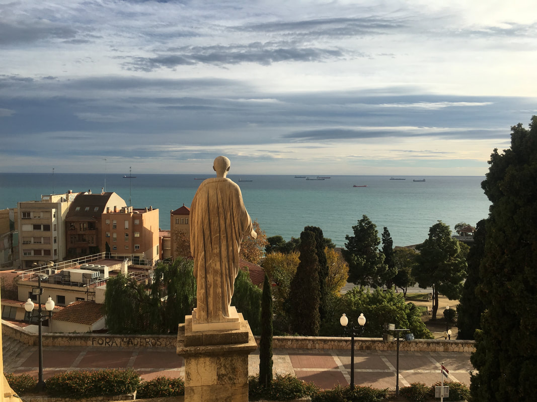
I also discussed ongoing collaborative work on the taphonomy of some Neandertal fossils from Poland. The work is ongoing, so more will be said about this topic when the research is complete. I officially began work today as a postdoctoral research at IPHES (Institut Català de Paleoecologia Humana i Evolució Social) in Tarragona, Spain. For the next two years I will be working on the "IDENTITIES" Project through a Marie Skłodowska-Curie Individual Fellowship (H2020-MSCA-IF-2016 No. 749188). I will continue to update this blog throughout the duration of my fellowship. Until then, enjoy these images taken from my new neighborhood.
|
John C. Willman
A place to find updates about my research. Archives
July 2021
Categories
All
|

 RSS Feed
