
|
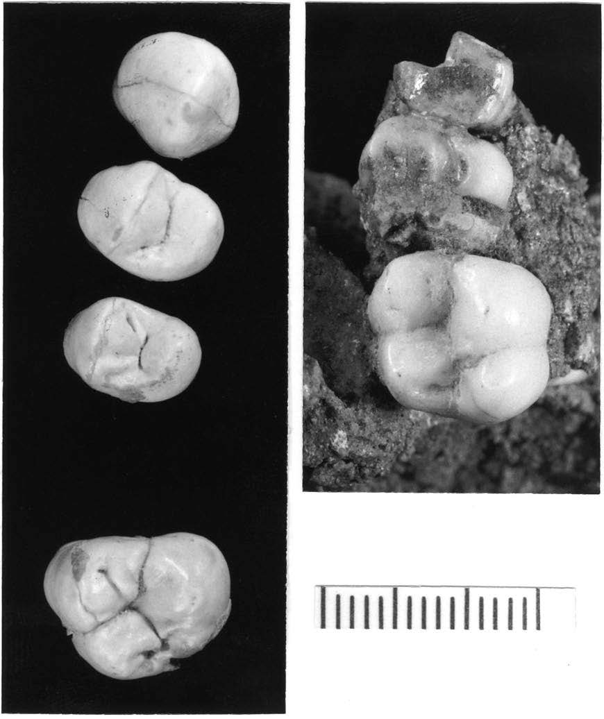
During the first two weeks of July, I had the opportunity to visit the Instituto Superior Técnico (IST) to microCT some Pleistocene human fossils from the sites of Gruta da Aroeira and Galeria de Cisterna. Both archaeological sites are located in the Almonda karstic system in Torres Novas, Portugal. My research stay was hosted by Prof. Manuel F. C. Pereira and Prof. António Maurício in the Laboratório de Mineralogia e Petrologia (LAMPIST) in the Centro de Recursos Naturais e Ambiente (CERENA) at IST where the microCT is housed. 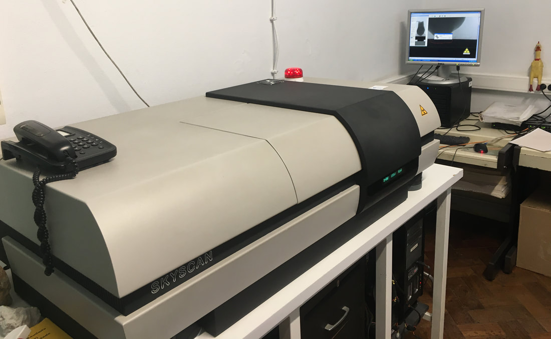 Above: MicroCT we used to scan the fossils. Yes, that is a rubber chicken in the background. Gruta da Aroeira is probably best known for the recent discovery of a ~400,000 year old (Middle Pleistocene) human cranium (Aroeira 3), but there are also two teeth (Aroeira 1 and 2) from older excavations that were published back in 2003. New analyses will focus on virtual approaches using the microCT scans we just produced. Analyses of internal tooth morphology will supplement what is already known from the earlier study of external morphology of these two teeth.
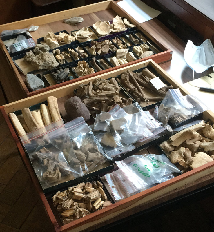
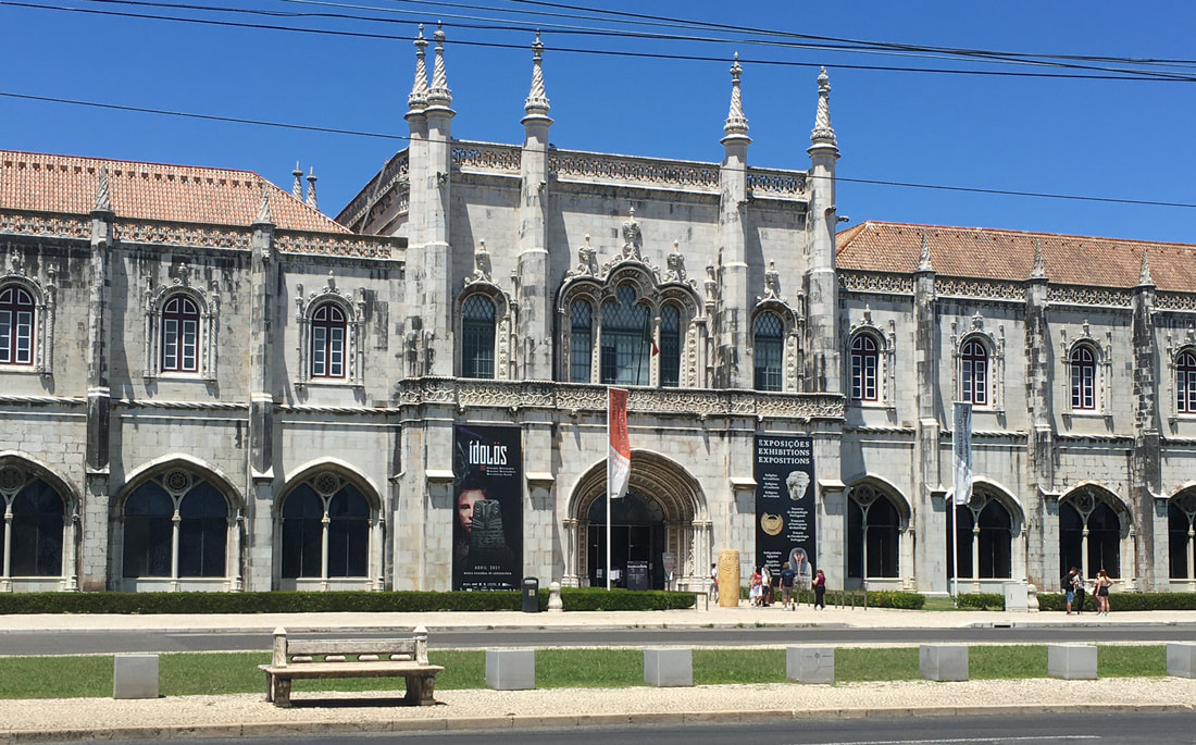 Above: Entrance to the Museu Nacional de Arqueologia where the fossils from Aroeira and Cisterna are curated. The temporary exhibit on "Idols" is fantastic. There are very few Pleistocene human fossils from Portugal, and only some of them have been examined using virtual anthropological approaches so far. Relevant comparative data from Portugal is a Neandertal tooth from Gruta da Oliveira (also part of the Almonda karstic system) and the dentition from the Gravettian 'Lapedo Child' (Lagar Velho 1). The VAPP Project will also include analyses of human fossils from the Gruta do Caldeirão (Tomar) dated to the Solutrean and Magdalenian. 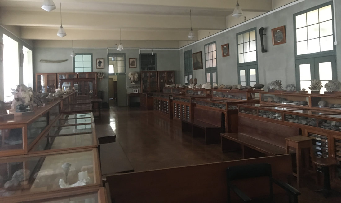 Above: A photo of the Museu Décio Thadeu—one of the geosciences museums at IST curated by Prof. Pereira.
References and further reading:Gruta da Aroeira
0 Comments
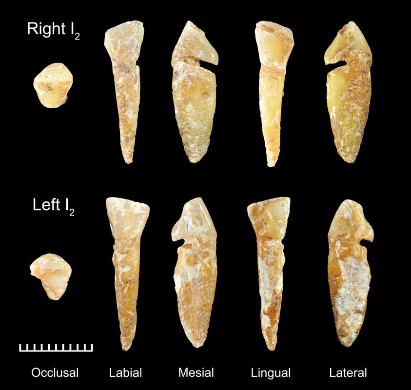 Above: Monte do Vale do Ouro 2 mandibular lateral (second) incisors. Large grooves are visible along lingual surface just below the crown. Scale increments are 1 mm.
The interpretation of the grooves was rather straightforward, since I was already familiar with some great previously published images—one featured the iconic photograph of "The Spinner" by Eugene Smith from a 1951 issue of LIFE magazine. I did a little searching of my own and was surprised to find a wealth of additional examples in the form of portraits and images in various publications from the 1800's and early 1900's wetting and hand spinning flax fibers (see below). This behavior, or similar behaviors relating to the manipulation of fibers, yarn, cordage, etc. across the surfaces of teeth provide good analogies for what may have created the deep grooves on the prehistoric teeth from Monte do Vale do Ouro 2. On a side note, it was fun to cite some artwork and the Brothers Grimm fairy tales! 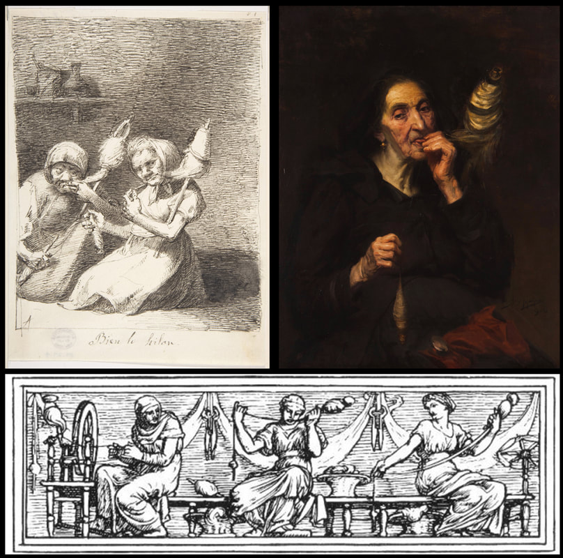 Above: Examples of flax wetting and hand spinning depicted in popular culture. Top left: Bien lo hilan (“They spin well” [1807-1845]) by Leonardo Alenza y Nieto (https://www.metmuseum.org/art/collection/search/334953, CC0 1.0). Top right: Velha Fiando (“Old Spinner” [1904]) by José Malhoa (https://commons.wikimedia.org/wiki/File:Velha_Fiando_(1904)_-_Jos%C3%A9_Malhoa.png, Public Domain). Bottom: The Three Spinsters (1886) by Walter Crane in Household Stories by the Brothers Grimm (http://www.gutenberg.org/ebooks/19068, Public Domain). What I considered even more amazing was that Alice Bernardo of Saber Fazer had made videos of contemporary women demonstrating the process of wetting and hand spinning flax fibers (see below). These examples provide plausible analogies for the types of behaviors that may have contributed to the formation of the wear and grooves documented in the Bronze Age individual from Monte do Vale do Ouro 2. We are not proposing that we have direct evidence of flax spinning/wetting by any means, but it is quite likely that our case study illustrates a prehistoric example of using the teeth to manipulate some kind of pliable cordage, yarn, or fibrous material in ways that are similar to the historic and contemporary examples we cite. 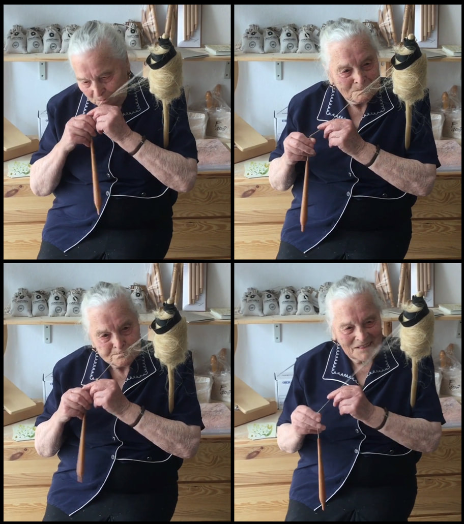 Above: Sequence of video stills of a woman spinning flax, approximately 3-4 seconds. Note slightly oblique, mediolateral guiding of flax fibers. Stills originate from video by Alice Bernardo (https://www.saberfazer.org/), used with permission. The original video can be streamed here. 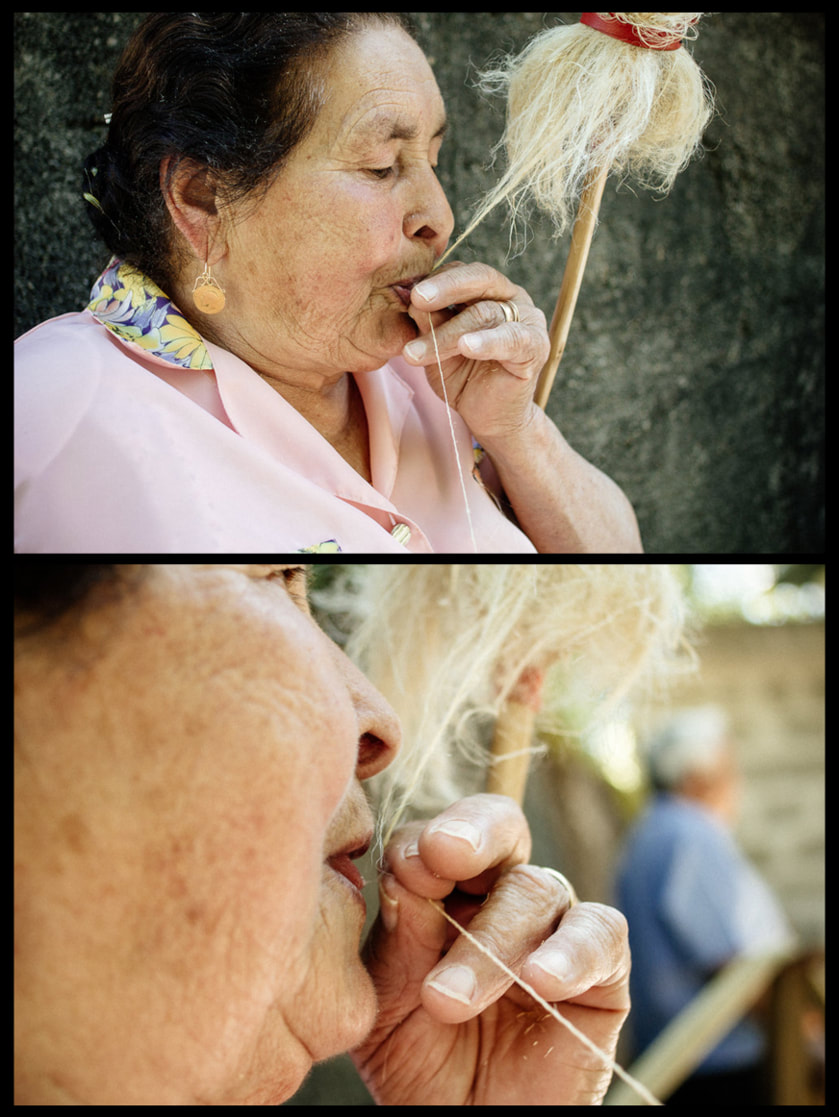 Above: Woman wetting flax fibers while hand spinning. Photographs by Alice Bernardo (https://www.saberfazer.org/), used with permission. The original video can be streamed here. The take away from the study of the two Bronze Age teeth from Monte do Vale do Ouro 2 is that they represent the embodiment of past human behaviors related to craft production. The depth of the grooves indicates that the individual also engaged in the behaviors that formed the grooves over a considerable period of their life. While there was an absence of grave goods, and not much other archaeological context to use to understand the life of this individual, these two little teeth provide significant insights into the social identity and lifeways of at least one individual from Monte do Vale do Ouro 2. References and further reading: Special note of thanks: Alice Bernardo of Saber Fazer allowed the use of images taken from videos she produced of the two women ("fiandeiras") spinning flax in the pictures above. I am extremely grateful for the privilege to use these images. The Saber Fazer website is truly a wonderful resource for those interested in traditions of craft production—especially archaeologists!
This study:
I had the opportunity contribute to two articles for a special issue of Quaternary International entitled "Not Only Use" that was edited by Juan Luis Fernández-Marchena, Lena Asryan, Antonella Pedergnana, and Andreu Ollé. The issue contains an impressive overview various methodologies used to study wear the burgeoning interest in multidisciplinary efforts to study wear traces (i.e., traceology) of different origins and the processes (i.e., operative chains) involved in manufacture, use, and abandonment of an object that is later recovered by an archaeologist (check out the editorial by Fernández-Marchena et al. for more info). The first article was lead by Raquel Hernando. In short, Hernando was interested in understanding if recent advances in optical microscopy could resurrect its use for the study of human dental microwear. Hernando noted that dental microwear analyses originally used optical microscopy, but it was later replaced by scanning electron microscopy (SEM). Eventually confocal microscopy became the favored technology for occlusal dental microwear analysis (i.e., dental microwear texture analysis, or "DMTA"), while SEM is still preferred for buccal microwear analysis. However, Hernando and colleagues note that SEM analyses are costly (we generally pay by the hour to use these microscopes) and the postprocessing of images is also quite time consuming (and exhausting for your eyes!). DMTA is generally quicker, but the microscopes—and software needed for DMTA—is not nearly as widely accessible as SEM. That means costly travel, lodging, and user fees to do DMTA analyses for many of us without local access to equipment. So, why not revisit optical microscopy? Hernando and colleagues point to many advances in optical microscopy that have been explored in the context of traceology. Buccal microwear seemed like the best place to start since it still widely uses SEM. Hernando and colleagues found that OM produces very similar results to traditional SEM methods whether one studies the original tooth or a dental cast of a tooth (see image below). 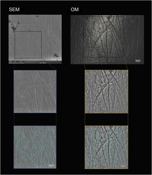 Above: Comparison of scanning electron (SEM) and optical microscopy (OM) images of buccal microwear. Note the excellent resolution in OM. Raquel Hernando and colleagues noted that optical microscopy provides many other advantages over traditional SEM analysis: less expensive equipment with less associated maintenance, wider accessibility of optical microscopes for researchers, less eye fatigue, greater image resolution, 3D appearance of images with greater definition, and relatively quick data acquisition and analysis. A drawback is the need to build up open-access databases for comparitive purposes, but the data produced in this paper marks the beginning of that effort. This study points out the importance of revisiting methodologies with a critical eye, but also how interdisciplinary research—something IPHES takes great pride in—can lead to innovation in allied fields of research. The second article explored the use of gigapixel-like (GPL) images for studying external surfaces of teeth. GPL images make use focus-stacking (extended focus images) and panoramic stitching of microscopic images to create mosaic images with high depth of field using SEM. This “gigapixel-like” (GPL) imaging strategy can be used to create multiscale, high-resolution images of entire, or partial, dental surfaces that can be viewed from a field of view that encompasses an entire tooth surface to high magnification views of dental microstructure, microwear, taphonomic features, among other features. The images have a variety of uses from the communication of results in scientific publications to their use in interactice museum displays and websites or training researchers. 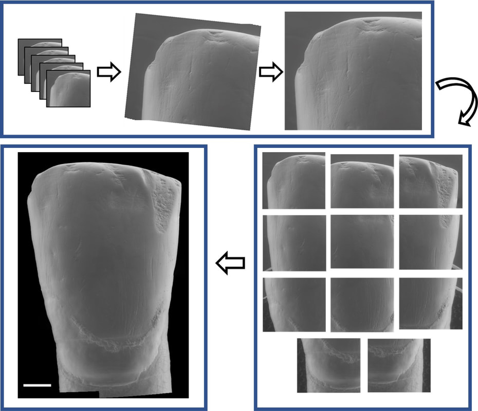 Above: simplified outline of focus-stacking and creation of image mosaic to create a gigapixel-like (GPL) image. 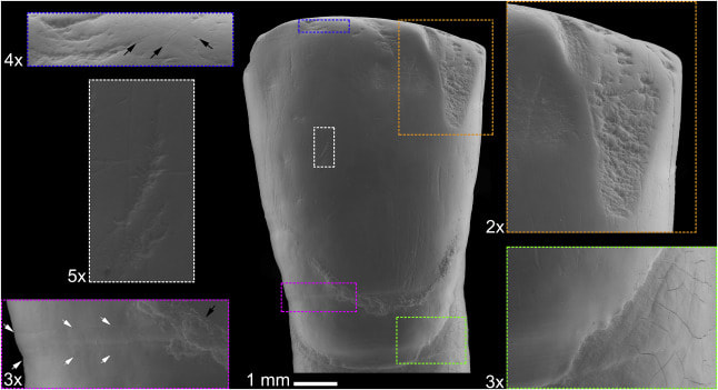 Above: A GPL image (center) with call-out boxes of varying magnification that indicate different surface features. Descriptions proceed clockwise from upper right corner. Orange rectangle: Medium size antemortem enamel chip with well-worn margins. Green rectangle: Detail of cementoenamel junction and root surface. Subtle perikymata (bottom left quadrant) and striations (upper left quadrant) are visible on the enamel. Subtle postmortem cracking of root surface also evident. Magenta rectangle: Detail of furrow-form hypoplasia with clearly visible perikymata (between white arrows). Black arrow points to dental calculus deposit. White rectangle: Detail of instrumental striation with a right oblique orientation. Blue rectangle: arrows indicate microstriations on labioincisal edge and a well-worn, but small, antemortem enamel chip to the left of the image. While the goal of the publication was to outline the GPL methodology and uses, we also made an interesting discovery from the creation of a GPL image for one of the teeth from the Chalcolithic context (dated to about 4000 years before present) of El Mirador Cave near Burgos, Spain. We found that at least one tooth exhibited a strange discoloration when viewed with the naked eye (see photo below). Microscopic examination revealed that the discoloration is related to enamel erosion—something that is rarely documented in prehistoric contexts. 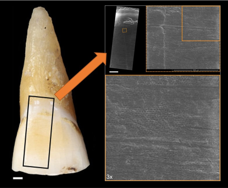 Above: Photo of original tooth with discolored (yellowish) enamel surface. GPL image sampling indicated by black box and GPL image indicated by orange arrow. Zooming in on section 300x shows "honey-comb" appearance of enamel surface. This indicates erosion of the enamel. This study makes me suspect that erosion in teeth from archaeological contexts is much higher than we currently acknowledge, and calls for a need for detailed analyses of the original teeth in conjunction with high magnification analysis for definitive diagnosis. Nonetheless, this is a very interesting (and rather accidental) discovery. More analyses of the El Mirador material are underway. References and further reading These studies:
Additional references:
There is a deep history of archaeological investigation focusing on the Bronze Age El Argar, or Argaric, cultural phenomenon from southeastern Spain. Argaric archaeology is probably most famous for the elaborate settlement structures, well-preserved burials, and evidence for sophisticated metallurgy and material culture. The rich archaeological record and excellent preservation of human remains have provided archaeologists with incredible resources for reconstructing the lifeways of these Bronze Age peoples.
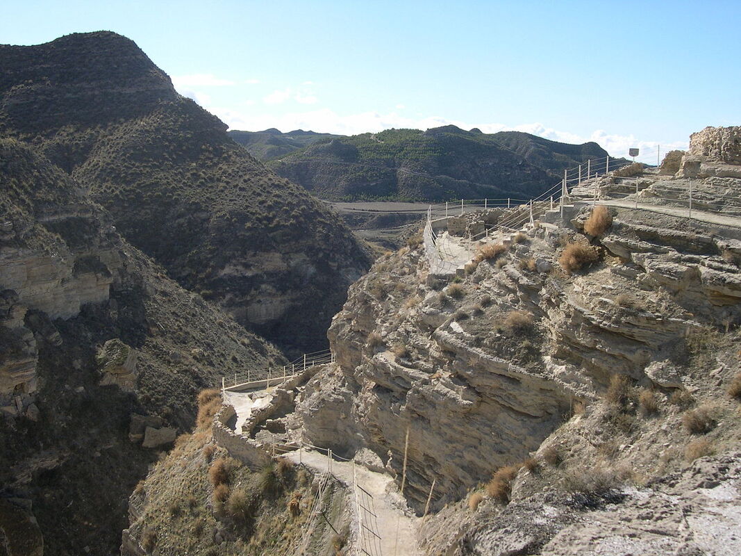
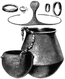
Above Left: view of the site of Castellón Alto. By Rafael Jiménez from Córdoba, España - Castellon Alto 2, CC BY-SA 2.0, https://commons.wikimedia.org/w/index.php?curid=21664792. Above Right: examples of grave goods recovered from an Argaric context. By Luis Siret - Siret, H., and Louis Siret (1887). «Les premiers âges du métal dans le sud-est de l'Espagne». Anvers., Public Domain, https://commons.wikimedia.org/w/index.php?curid=1294319 I was recently involved in a collaborative research project on the human remains from the site of Castellón Alto that focused on the dental remains of the individuals buried at the site. As part of his Ph.D. research on the human remains from the site, Àngel Rubio discovered an interesting trend at the site: of the 106 burials examined, the teeth of 5 individuals showed atypical patterns of dental wear (see below). What was even more astounding was that each of those individuals was female. No males had these interesting patterns of wear. Further microscopic analysis conducted by Dr. Marina Lozano provided clues as to what behaviors may have contributed to the unique wear patterns identified on the teeth of these 5 individuals. 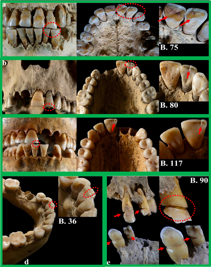 Above: The 5 female individuals with atypical patterns of dental wear. Red arrows and circles indicate the location of the wear in the photos. Image from the article: https://doi.org/10.1016/j.jas.2020.105239. The remarkable preservation of organic remains (textiles, wool, plant fibers, etc.) in addition to extensive durable material culture (awls, loom weights, spindle whorl, needles) found at Argaric archaeological sites provided additional clues as to what tasks may have contributed to the unique patterns of dental wear on the 5 women from Castellón Alto. A probable explanation is that at least some of the women at the site were involved in specialized craft production such as textile production, processing of fiber or cordage, basketry, and similar tasks (see illustration below). Ethnohistoric documentation of the use of the teeth for craft production adds additional support that the formation of atypical dental wear in the subset of the women from Castellón Alto was related to craft production. 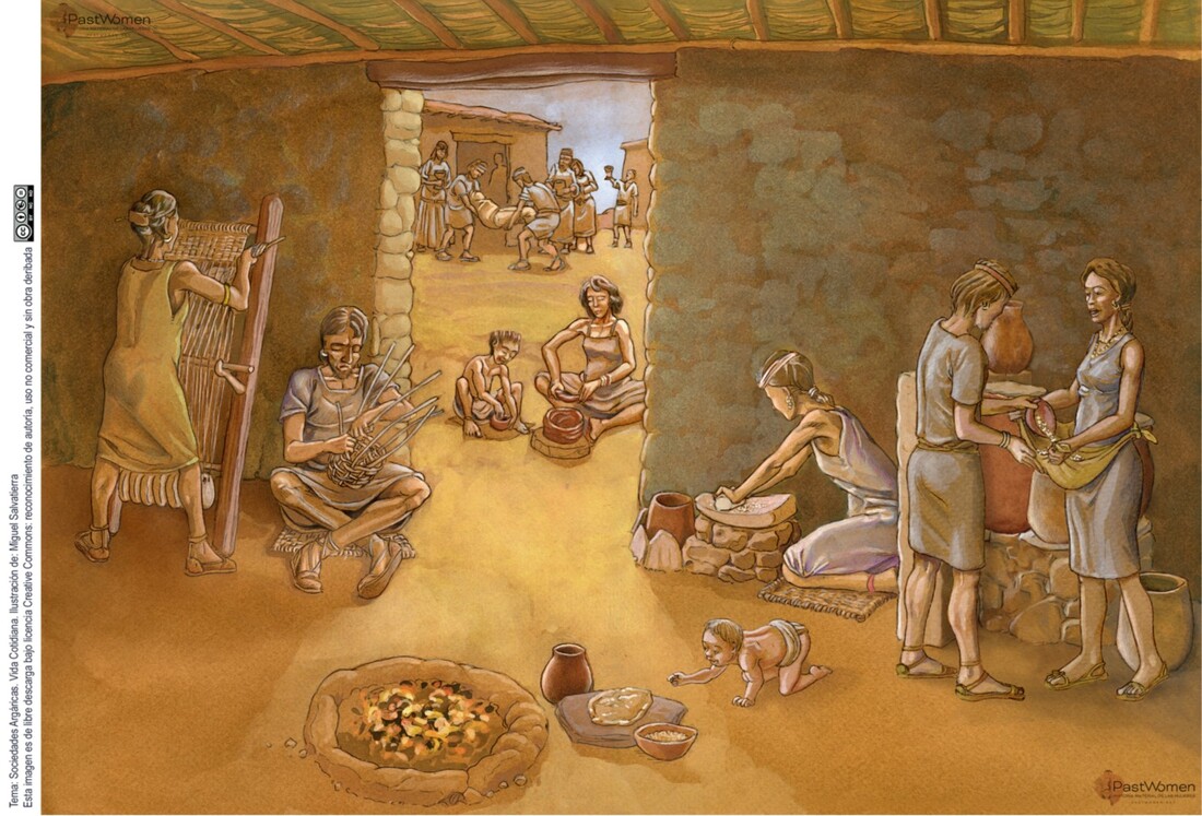 Above: A scene of Argaric life featuring the many of the tasks related to food preparation and craft-production in the foreground. Ilustración: Miguel Salvatierra "Cultura argárica". The rich archaeological record from the Argaric contexts of southeasten Spain is bound to reveal more insights into human social lives and identities of Bronze Age peoples. In this case, analyses have revealed a unique role for at least some of the women buried at the site engaged in. References and further readingThis study:
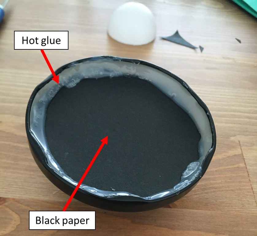
Lozano M, Jiménez-Brobeil SA, Willman JC, Sánchez-Barba LP, Molina F, and Rubio Á. 2020. Argaric craftswomen: Sex-based division of labor in the Bronze Age southeastern Iberia. Journal of Archaeological Science:105239. https://doi.org/10.1016/j.jas.2020.105239. Additional references: I must emphasize the great resources at http://www.pastwomen.net/ and the resources on the Argaric culture in particular (http://www.pastwomen.net/objetos/cultura-argarica) for the preparation of this post. The website offers incredible, multi-language resources for the instructors, scholars, and anyone that is curious about the social lives of women in the past. The researchers, artists, and other contributors have really created an incredible resource. I use a lot of photography and microscopy to document minute surface details on the fossil and bioarchaeological human remains I study. A problem I constantly deal with is getting the surface I want to visualize in the appropriate orientation—especially if the object is small and fragile (e.g., teeth). Some researchers use small sandboxes, and this is effective, but it presents a few problems. Abrasion from sand is probably the biggest concern, since it can cause taphonomic wear--a real problem for those of us interested in using dental wear for dietary and behavioral reconstructions. Black aquarium sand is often preferred, but it is not only abrasive, but so fine that it is often difficult to remove from the surfaces of the object you place in it when you are finished photographing them. Lots of researchers like to use modeling clay, but clay is also extremely difficult to remove from surfaces. Even if a surface looks clean, you are bound to see some clay residue under high magnification. Some researchers also have a tendency to push the object they are studying into the clay which can damage fragile artifacts or wedge clay into tiny crevasses and cracks. The more time you send adjusting the object for imaging introduces more opportunities to damage, abrade, or dirty the artifact you are trying to document. A good solution for small objects is to use a tilting stage that allows you to orient the object without touching it over and over again. I have wanted a microscope tilting stage for quite some time, but they are generally not cheap when you do come across one. I did a bit of searching and came across a DIY stage with step-by-step instructions online, and decided to try my luck at making something similar with objects I have in my apartment and a couple of small purchases. The tools I used (hot glue gun and hobby knife) are easy to come by, and the supplies I purchased (a washer, small screw nuts, black paper, and ping pong balls) are super cheap and easy to acquire. I spent about 2 euros and found the rest of the supplies sitting around my apartment. 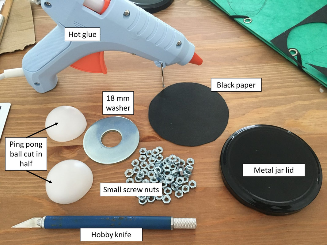 You can probably substitute a number of other household objects and still make a similar tilting stage. The ideal "nuts" would be lead shot, fishing line weights, etc. You could probably use a racquetball in place of the ping pong ball like my colleagues do at IPHES, but you may need to swap out the washer base for something larger (e.g., a section of pvc tubing, thick-walled poster tube, etc.). I have been playing around with the idea of using a magnetic ball mount kit intended for cell phones, but this DIY project was easier and quicker than ordering stuff online. 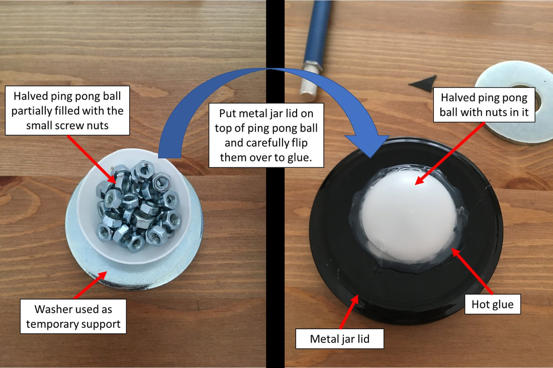 Play around with the ping pong ball a little and adjust the number of nuts used as counter weight if needed. My glue gun runs hot but still didn't melt the ping pong ball. Be careful when gluing the ball to the lid because the lid will get hot! I pressed down lightly on the ping pong ball to ensure a good seal as the glue dried.
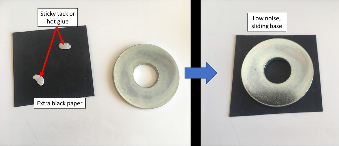 The washer made a lot of noise when I place the finished tilting stage on my portable microscope stand. I decided to stick a piece of paper on the bottom to prevent it from scratching the stand base and reduce the noise. I generally use a piece of black paper under whatever I am viewing to help slide the object around under the microscope (it's a lot easier than using the x-y adjustment stage I have when working at low magnification anyway). The tilting stage slides around effortlessly on my microscope stand with the added paper. 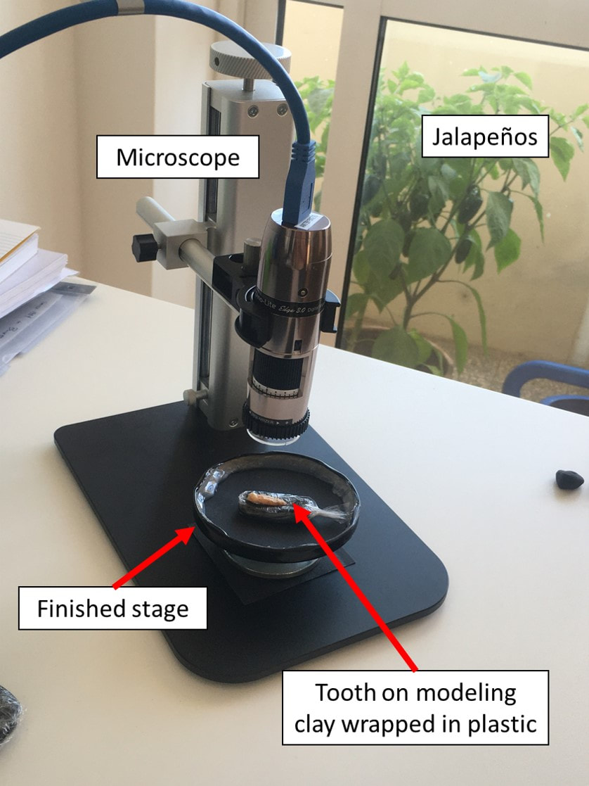 The entire process took less than 10 minutes and 2 euros to complete. Plus, I still have 4.5 ping pong balls left... I'll probably modify this stage or build different stages as I come across better supplies in the future. I will make a new post if I make any significant updates. For now, this is a very efficient tool for imaging loose teeth with a portable microscope and digital macrophotography, and I am very happy with the result.
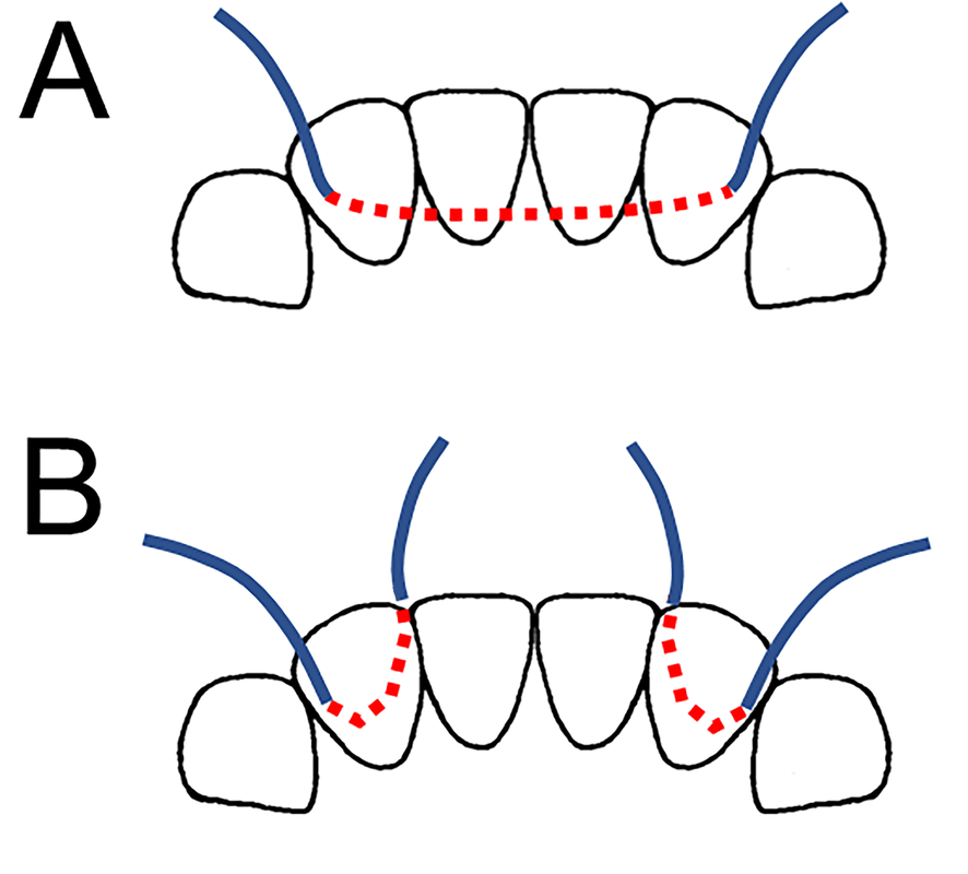
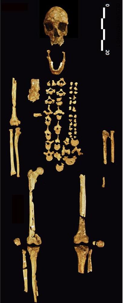
Ohalo 2 is a 23,000 year old hunter-fisher-gatherer that was recovered from a burial at a spectacularly well-preserved archaeological site on the shore of the Sea of Galilee. In 2013, I visited Tel Aviv University to collect data on the human fossils curated in the Department of Anatomy and Anthropology for my doctoral dissertation. While examining the Ohalo 2 skeletal material, I noticed, the missing upper central incisor and a fully healed socket. The teeth immediately surrounding this incisor were present - although a few neighboring teeth were broken postmortem (not surprising for a fossil of this antiquity). The dental wear in the upper jaw was fairly typical of a Late Pleistocene hunter-fisher-gatherer; but when I picked up the mandible, I noticed that the lower incisors had an interesting curvature to their wear plane. This modest "arch-like" pattern of dental wear perfectly matched the position of the missing upper incisor. The overall pattern of dental wear and healed socket was starting to look a lot like the intentional tooth removal - or "ablation" - something that had not been suggested in previous analyses of Ohalo 2. Interestingly, the Natufians - the archaeologically-defined culture that existed some 10,000 years after Ohalo 2 - practiced dental ablation. Could there be a biocultural connection between this Early Epipaleolithic forager and the Late Epipaleolithic Natufians? Ten-thousand years is quite a leap in time, but long-term continuity in some aspects of Epipaleolithic human behavior in southwest Asia had been suggested based on other aspects of the archaeological record. 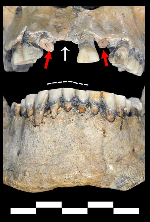 Above: The white arrow points to the tooth that was lost antemortem (in life) as indicated by the healed bone and resorbed socket. The dotted white line shows the uneven plane of wear that indicates that the individual was alive long after the tooth was removed, since dental wear continued to progress - albeit unevenly due to the lack of an occluding upper right central incisor. Red arrows show teeth that are broken and missing postmortem. My colleague, Dr. Sarah A. Lacy, also had the opportunity to study the Ohalo 2 skeletal material in Tel Aviv a few years before me, so we decided to collaborate on an assessment of the Ohalo 2 oral pathological conditions. Our thinking was that a differential diagnosis of the tooth loss may help us understand whether the incisor was lost through the intentional cultural practice of dental ablation or by some other cause. Given the small number of fossils from the Eastern Mediterranean that date to this time period, we also thought it would be an important source of comparative data for researchers interested in Late Pleistocene human paleobiology and behavioral reconstructions. 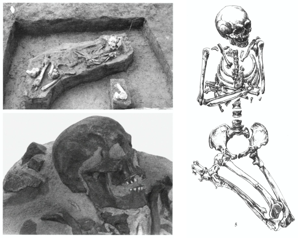 Above: The burial of Ohalo 2 during excavation, a close-up of cast of individual's cranium, and an drawing of the skeletal elements after excavation in laboratory. Image modified from: https://www.jstor.org/stable/41492570. In addition to the loss of the upper central incisor, we found a number of other pathological conditions: two carious lesions ("cavities"), pulp exposure, and mild to moderate bone loss around the front teeth. Another cool finding is the probable agenesis of the left upper third molar - "agenesis" means that the tooth never developed at all. We noted that these conditions are not really abnormal for a Late Pleistocene human, but the general state of oral health did not seem to contribute to the loss of the upper incisor. So, if the other oral pathological conditions were unlikely contributors to the loss of the incisor, what about loss through a traumatic event? A bad fall, interpersonal violence, or some other kind of accident could have knocked out Ohalo 2's front tooth. At first, we thought trauma was a good candidate since Ohalo 2 has an absolutely astonishing bony growth in their chest. The growth was due to infection (chronic osteomyelitis) caused by localized trauma to the lower thorax. Could the incisor have also been knocked out at the same time? Possibly, but unlikely. We determined that the tooth was more likely lost in early in life (childhood or early adolescence) by examining factors related to the timing of tooth eruption and expectations regarding progressive dental wear in hunter-gatherers. In contrast, the major thoracic injury was more likely a later life occurrence - and a rather debilitating one for Ohalo 2. Intentional tooth removal - "ablation" - seems to be the best candidate for Ohalo 2's lost incisor. The tooth was probably lost around late childhood/early adolescence - an important period of physical and social maturation for a young person. Indeed, it is around this age that body modification practices often occur, as shown by numerous other examples documented from bioarchaeological and ethnohistoric contexts. 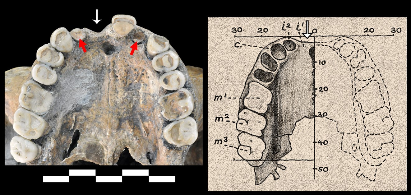 Above Left: The occlusal view of the maxillary (upper) teeth of Ohalo 2. The white arrow is pointing to the tooth that was lost antemortem (before death). The bone is resorbed/healed. The red arrows point to teeth that were lost postmortem. Above Right: A line drawing of a Natufian individual from Shukbah showing the empty space (white arrow) where a tooth was lost before death in the exact location that is seen in Ohalo 2. Photograph by JC Willman. Line drawing modified from Keith 1931. Since the Natufians also practiced incisor ablation, and many behavioral trends among the Natufians seem to have deep antiquity within the Epipaleolithic of the region, we thought it was interesting to also see this biocultural practice portrayed by Ohalo 2 as well. With so few fossils dated to this time period in southwest Asia, it is hard to say whether or not this is truly a biocultural trend spanning the Early and Late phases of the Epipaleolithic, but it is an exciting possibility. Hopefully, future discoveries will shed more light on the antiquity and patterning of Epipaleolithic body modification practices in southwest Asia. Until then, Ohalo 2 represents one of the earliest probable cases of dental ablation in western Eurasia, and more evidence for the embodiment of human social identities through intentional body modification practices among Late Pleistocene peoples. References and further reading:This study:
Access the paper for free until 19 July 2020 here: https://authors.elsevier.com/a/1b98C6hTLQdsfi Willman JC, and Lacy SA. 2020. Oral pathological conditions of an Early Epipaleolithic human from Southwest Asia: Ohalo II H2 as a probable case of intentional dental ablation. International Journal of Paleopathology 30:68-76, https://doi.org/10.1016/j.ijpp.2020.04.001. Additional references: Bocquentin F. 2011. Avulsions dentaires et identité régionale chez les Natoufiens. Tüba-Ar (Turkish Academy of Sciences Journal of Archaeology) 14:261-270. Link Bocquentin F, Crevecoeur I, and Semal P. 2013. Artificial modification of the central upper incisors of Homo 4 (Plot XX J burial). In: Edwards PC, editor. Wadi Hammeh 27, an Early Natufian Settlement at Pella in Jordan. Leiden: Brill. p 383-387. Link Edwards PC. 2015. Natufian interactions along the Jordan Valley. Palestine Exploration Quarterly 147(4):272-282, doi.org/10.1179/1743130115Y.0000000001. Hershkovitz I, Edelson G, Spiers M, Arensburg B, Nadel D, and Levi B. 1993. Ohalo II man—unusual findings in the anterior rib cage and shoulder girdle of a 19000‐year‐old specimen. International Journal of Osteoarchaeology 3(3):177-188, https://doi.org/10.1002/oa.1390030304. Hershkovitz I, Speirs MS, Frayer D, Nadel D, Wish-Baratz S, and Arensburg B. 1995. Ohalo II H2: A 19,000-year-old skeleton from a water-logged site at the Sea of Galilee, Israel. American Journal of Physical Anthropology 96(3):215-234, http://dx.doi.org/10.1002/ajpa.1330960302. Keith A. 1931. New discoveries relating to the antiquity of man. London: Williams & Norgate. Link Maher LA, Richter T, and Stock JT. 2012. The pre-Natufian Epipaleolithic: long-term behavioral trends in the Levant. Evolutionary Anthropology 21(2):69-81, https://doi.org/10.1002/evan.21307. Nadel D. 1994. Levantine Upper Palaeolithic–Early Epipalaeolithic burial customs: Ohalo II as a case study. Paléorient 20(1):113-121, www.jstor.org/stable/41492570. Snir A, Nadel D, Groman-Yaroslavski I, Melamed Y, Sternberg M, Bar-Yosef O, and Weiss E. 2015. The Origin of Cultivation and Proto-Weeds, Long Before Neolithic Farming. PloS one 10(7):e0131422, https://doi.org/10.1371/journal.pone.0131422. Trinkaus E. 2018. The palaeopathology of the Ohalo 2 Upper Paleolithic human remains: A reassessment of its appendicular robusticity, humeral asymmetry, shoulder degenerations, and costal lesion. International Journal of Osteoarchaeology 28(2):143-152, https://doi.org/10.1002/oa.2640. The last few months have been a bit chaotic to say the least. I wrapped things up at IPHES in December, packed up my belongings, and drove across the Iberian Peninsula with my partner. January 6, 2020 marked my first day on a new MSCA-IF postdoctoral research project at the Universidade de Coimbra. I joined CIAS - Research Centre for Anthropology and Health (Centro de Investigação em Antropologia) - in the Department of Life Sciences (Departamento de Ciências da Vida) at the University of Coimbra where I am working with Ana Maria Silva in the Laboratory of Prehistory. I will be studying prehistoric human dental morphology with a focus on Portuguese archaeological sites spanning the Paleolithic to Bronze Age over the next two years. The project will incorporate methods of assessing external crown and root morphology as well as virtual approaches to incorporate internal and three-dimensional dental morphology. Given the focus on virtual approaches, we named the project "Virtual Anthropology of Prehistoric Portugal" or VAPP for short. I'll continue using this blog to provide updates on the VAPP Project, as well as news of research coming out of the IDENTITIES Project and other collaborations. "Why Portugal?" Last week I had my first Portuguese language class in Coimbra. There were general introductions from the students and some follow-up questions from our instructor. When it was my turn, I introduced myself as being from the US and working at the University of Coimbra as a biological anthropologist studying archaeological human remains. I usually get a lot of puzzled looks and follow-up questions when I introduce myself like this, but that wasn't the case this time. Instead, I was asked, "Why Portugal? Why Coimbra?" My short answer was that I came to Portugal as an undergraduate, fell in love with the country, and have been wanting to come back ever since. I've been reflecting on this a bit lately because I never really thought that I would live in Portugal, just come back and visit once in a while. Even more inconceivable to me on reflection is that I have now lived on both sides of the Iberian Peninsula! But why Coimbra?I had a bunch of meetings during my first postdoc at IPHES that were focused on documenting the progress of the IDENTITIES Project but also on professional development. Among the most important meetings was an early one I had with Ignasi Pastó about research trajectories in Europe. Ignasi encouraged me to think about applying for another Marie Curie fellowship, and at the end of the meeting he said, "I will follow up with you in May [2018]. You should have an idea of what you want to work on and who you want to collaborate with." So, I was one and a half months into a new postdoc and already planning the next one! My pipe dream of moving to Portugal resurfaced at that moment. I also knew that I wanted to gain greater knowledge of dental morphology and virtual approaches to human remains. So, I sought some advice from trusted colleagues about collaborators in Portugal and they kept recommending CIAS and the Laboratory of Prehistory at the University of Coimbra. So, I got in contact with Ana Maria Silva and proposed the collaboration that would become the VAPP Project. Long story short: we wrote a successful grant application and now I am in Coimbra. Yesterday, Jane Buikstra gave a talk at CIAS called "Bioarchaeology Goes Global & Historical." It was great to see a world-renowned bioarchaeologist give a talk, but it was even more incredible to hear her say that "the University of Coimbra has the best research institution in Bioarchaeology on the continent." So, if Jane Buikstra says so, I don't find it necessary to argue the point any further. Coimbra is a solid choice. Oh, pastries are also a good way to choose postdoc locations....  The practice of body modification - often related to the expression of individual or group-level social identity - is not uncommon in prehistory. However, body modification can be difficult to identify since so much of it is ephemeral or not likely to preserve in such a way that we can identity it as related to adorning or modifying the body. For instance, many forms of body modification lack archaeologically-visibility because they only affected soft tissues (body painting, hair style, ear and nose piercings, tattoos, scarification, etc.) which are only preserved in very specific cases (e.g., bog bodies, mummies). However, body modification that affects the human bone and/or teeth of once living people is more likely to preserve in the archaeological record. Examples include cranial shaping/modification, dental ablation (the removal of teeth for cultural/aesthetic reasons), intentional tooth filing or chipping, and facial piercings (labrets) that abrade the teeth. As a biological anthropologist specializing in dental wear, I have come across a lot of very interesting cases of intentional dental modification (e.g., ablation, chipping, filing) and some cases of unintentional dental wear - my favorite being that caused by the prolonged wearing of labrets. These cases always peak my interest because they are great opportunities to discuss aspects of human identity and lived experience in prehistoric contexts - something that is often quite difficult to elaborate on in Pleistocene contexts.
I enlisted the help of my colleague Raquel Hernando to analyze the dental wear, and together with Isabelle and Marie, we wrote up a manuscript for the American Journal of Physical Anthropology. An initial difficulty we faced was context. There is no reliable direct date for OH1 and many doubt an earlier date of 16,920 ± 920 BP. We got around this issue with a morphological assessment of biological affinity using the relatively complete OH1 mandible. We showed that OH1 mandible more closely matches the morphology of other Late Pleistocene and Early Holocene fossils from Africa, but is outside of the variation seen in recent humans. Earlier analyses of Late Pleistocene fossils (e.g., Ishango, Lukenya Hill) also show that aspects of cranial and postcranial morphology of OH1 follow a Late Pleistocene rather than recent human morphological pattern as well. So, the various morphological assessments provide good support for a Late Pleistocene age for the OH1 skeleton. While an exact date isn't possible, the morphological similarity to other Pan-African fossils from the Late Pleistocene suggests that OH1 dates to somewhere in the ballpark of 20,000-12,000 year ago. We then described the pattern of wear on the mandibular front teeth and the cheek teeth. We showed that the front teeth exhibit a pattern of wear that resembles the use of an large facial piercing, or labret, in the lower lip. We suggest that the more surprising facets on the cheek teeth correspond to labrets being worn through piercings in the cheeks. We concluded that OH1 likely wore three different facial piercings - one through the lower lip and one in each cheek. So far, this has never been described paleoanthropological or bioarchaeological contexts in Africa. This is exciting because it adds to the known diversity of body modification practices already documented in Late Pleistocene and Early Holocene Africa. The dominate pattern in the Terminal Pleistocene (20-12,000 years ago) is the ablation of teeth - especially in northeast and northwest Africa; but by the Early Holocene (10-7,000 years ago) some evidence for labret-use, in addition to ablation, begins to show up in the African archaeological record. By the Middle Holocene (7-3,000 years ago) we begin to see ablation, labret-use, and chipping/filing throughout Africa. This diversity of body modification practices is interesting because it may reflect the movements and interactions of prehistoric peoples through time and space. 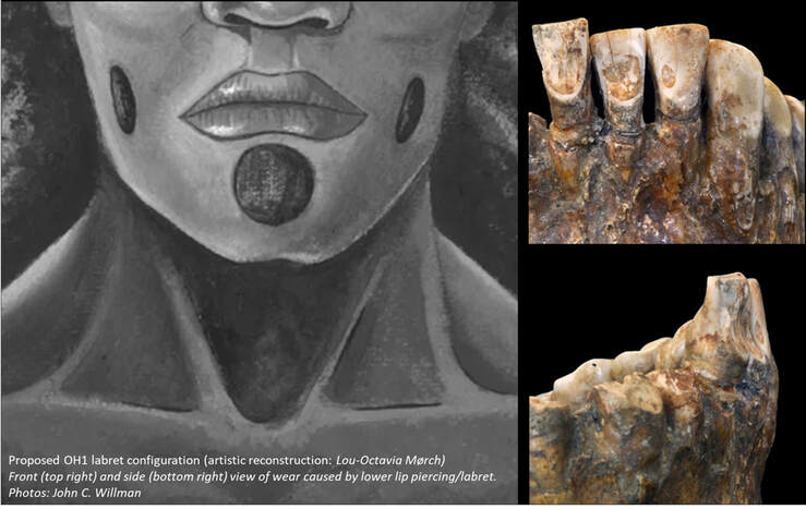 Above left: An artistic reconstruction (by Lou-Octavia Mørch) of the proposed pattern of labret piercings worn by OH1. Above rigt: the front and side view of the incisors and canines. The darker areas of enamel on the teeth is the exposed dentin that we propose was caused by a lower lip labret. Body modification often marks specific events related to social maturation (e.g., puberty, marriage, adult status, etc.) during an individual's life. This all means that labret wear on teeth of OH1 provides indirect evidence for personal adornment that is probably tied to social practices and individual or group identity. We hope that this research stimulates new research on human body modification and social identities in the Pleistocene as it has the opportunity to reveal a greater depth of understanding about the social lives of past peoples as more case studies are available for intra- and inter-regional comparisons. Further reading and references:This study:
Willman JC, Hernando R, Matu M, Crevecoeur I. 2020. Biocultural diversity in Late Pleistocene/Early Holocene Africa: Olduvai Hominid 1 (Tanzania) biological affinity and intentional body modification. American Journal of Physical Anthropology, doi.org/10.1002/ajpa.24007. Additional references: Crevecoeur I, Brooks A, Ribot I, Cornelissen E, and Semal P. 2016. Late Stone Age human remains from Ishango (Democratic Republic of Congo): New insights on Late Pleistocene modern human diversity in Africa. Journal of Human Evolution 96:35-57, http://dx.doi.org/10.1016/j.jhevol.2016.04.003. Matu M, Crevecoeur I, and Huchet JB. 2017. Taphonomy and Paleoichnology of Olduvai Hominid 1 (OH1), Tanzania. International Journal of Osteoarchaeology 27(5):785-800, http://dx.doi.org/10.1002/oa.2593. Parsche F. 1993. Peculiarities on the incisors in the mandible of the skull Olduvai I. HOMO 44(1):30-36. Tryon CA, Crevecoeur I, Faith JT, Ekshtain R, Nivens J, Patterson D, Mbua EN, and Spoor F. 2015. Late Pleistocene age and archaeological context for the hominin calvaria from GvJm-22 (Lukenya Hill, Kenya). Proceedings of the National Academy of Sciences, https://doi.org/10.1073/pnas.1417909112. I had the great privilege of contributing to a new article published in PLOS ONE yesterday that was coordinated by my colleague, Dr. Kristin Krueger, of Loyola University Chicago. The piece has been summarized nicely by Katherine J. Wu at PBS NOVA. I provide a short summary and a few additional thoughts here. Neandertal and early modern human anterior tooth-use Reconstructions of Neandertal behavior frequently call attention to their anterior tooth (incisor and canine) wear. Some incredible examples of extreme anterior dental wear are documented among some of the earliest well-published Neandertal fossils (for example: La Ferrassie and Forbe's Quarry) and the iconic photo by Erik Trinkaus of Shanidar 1's anterior dental wear (see below) is widely used to illustrate the phenomenon. This high degree of wear in the front teeth is most frequently interpreted as evidence that Neandertals used their "teeth-as-tools" or as a "third hand" regularly. In fact, it's often rare not to see a behavioral reconstruction of Neandertal that doesn't depict them using their "teeth as tools" in a museum display, artistic reconstruction, and television popular science documentaries. 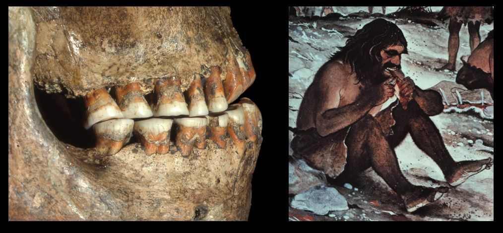 Above left: Shanidar 1 Neandertal with heavy anterior tooth wear (Photo: Erik Trinkaus). Above right: Artistic reconstruction of a Neandertal using its anterior teeth (as a "third hand") to clamp down on a piece of meat while they cut it into smaller pieces using a stone tool with their free hand. This behavior is also routinely called "stuff and cut" - a phrase that can be attributed to C. Loring Brace. 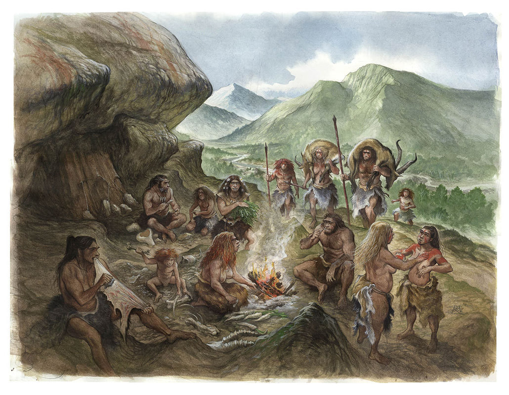 Above: Artistic reconstruction of a Neandertal group from El Sidron Cave featuring a Neandertal woman using her teeth to assist in hide scraping activities. Another behavior that leaves traces on teeth is the use of toothpicks which I discuss elsewhere. Artwork by Emmanuel Roudier. The robust build of Neandertal skulls and their large anterior teeth are a stark contrast to the smaller teeth and more gracile skulls of modern humans. These morphological differences were coupled with the observations of heavy anterior dental wear, and many researchers saw aspects of Neandertal cranial and dental morphology as adaptations that helped dissipate the high bite forces and/or repetitive loading of the anterior teeth and face when engaging in the use of teeth-as-tools. This "Anterior Dental Loading Hypothesis" has held a prominent position in Neandertal research. But why use your teeth as tools? The technological capacities of the Neandertals were (and often still are) considered impoverished or inferior to those of Upper Paleolithic early modern humans. Thus, early modern humans were thought to possess a degree of technological ingenuity that reduced morphological selection for the robust cranial and dental features found in Neandertals and their predecessors. The anterior dental loading hypothesis and ideas of "Neandertal inferiority/modern human superiority" have been challenged on a number of grounds. However, dental wear can provide direct evidence for how the anterior dentition was used among Neandertals and early modern humans. However, few studies directly compare Neandertals and early modern human dental wear, and none have used dental microwear texture analysis to test these ideas.  Above: The Shanidar 1 Neandertal (left) compared to an early modern human from Předmostí (right). Many cranial features, and the front teeth in particular, are quite large and "robustly built" in Neandertals but reduced in size and "gracile" in early modern humans. This is where our new paper jumps into the discussion. The research, coordinated by Dr. Krueger, uses dental microwear texture analysis of the surfaces of anterior teeth to explore similarities and differences in Neandertal and early modern human tooth-using behaviors. The results were surprising given the amount of previous research emphasizing behavioral and morphological differences between the two groups. We compared Neandertal and early modern human anterior dental microwear textures to each other as well as to a large comparative databases for human groups with considerable variation in temporal, ecological, geographic, and cultural backgrounds (see map below). While I have written elsewhere about how molar microwear texture can be used to reconstruct diets in Homo sapiens and Neandertals, the microwear textures on the anterior teeth are somewhat different. Incisors and canines are not only involved in the breakdown of food but are frequently used in a variety of non-dietary tasks. Therefore, dental wear on front teeth is a palimpsest of behavioral signatures related to dietary and non-dietary (using the “teeth-as-tools”) behaviors as well as various environmental factors (often related to the presence or absence of grit and dust). The excellent contextual information available for the human groups used for comparative purposes includes information on the diet, non-dietary behaviors, environment, and other factors that we use to interpret the results obtained for the Neandertals and early modern humans. 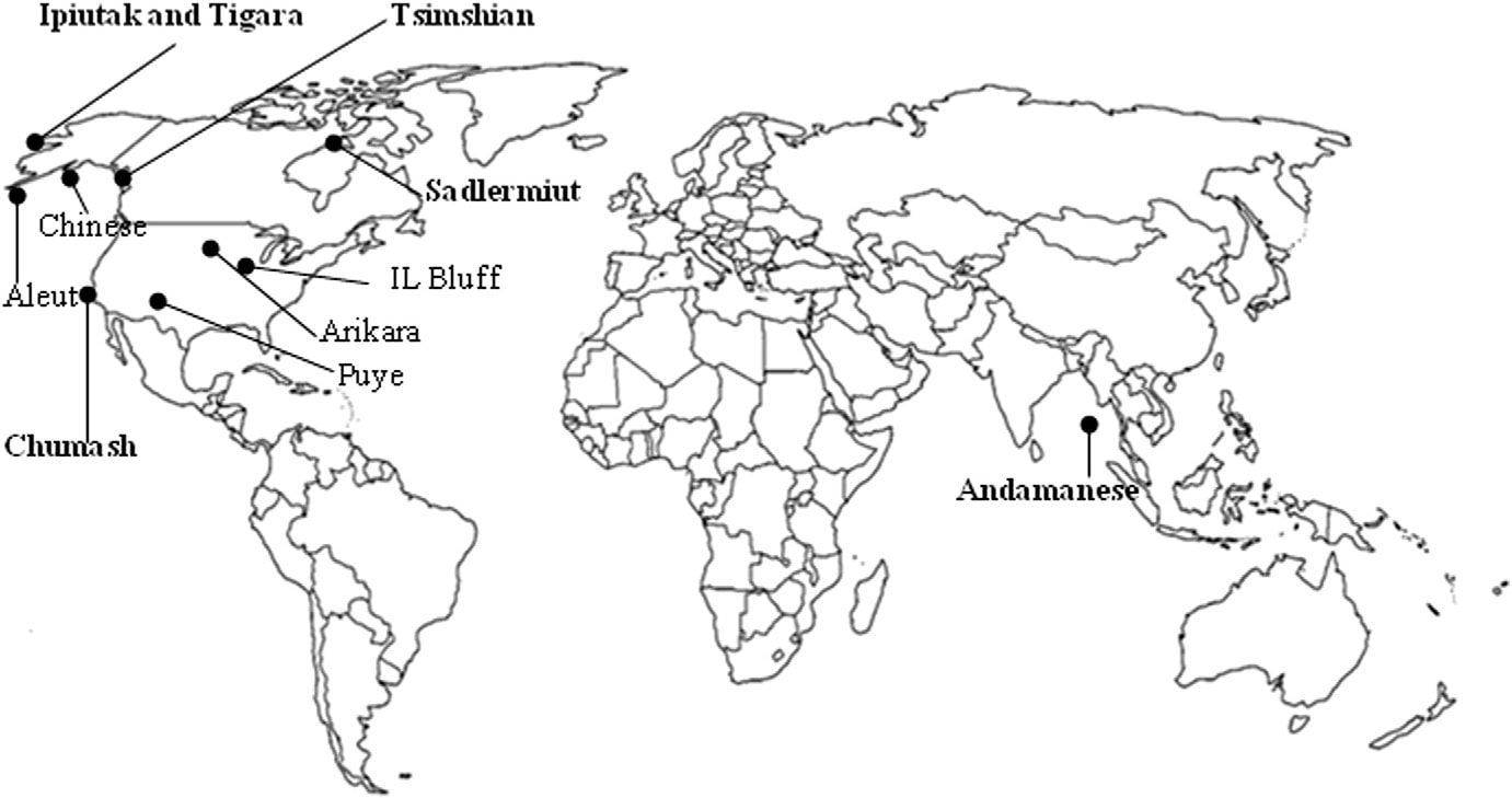 Above: Map showing the location of human groups used to compare and contextuale the dental microwear texture results for Neandertals and early modern humans. Map is from Krueger 2015. Interestingly, we found that the Neandertals and early modern humans exhibited very similar anterior dental microwear texture results. The values for both of these Pleistocene human groups were closest to those of the Point Hope Tigara hunter-gatherers from Alaska in our comparative sample. The Tigara are known to have used their anterior teeth in a variety of non-dietary, clamping and grasping behaviors such as as processing/softening animal hides and working sinew into thread. Thus, one way of interpreting the data for Neandertals and early modern humans were engaging in a similar range of clamping and grasping behaviors with the front teeth. Clothing and hide processing are reasonable behaviors documented in bioarchaeological and ethnohistoric contexts. 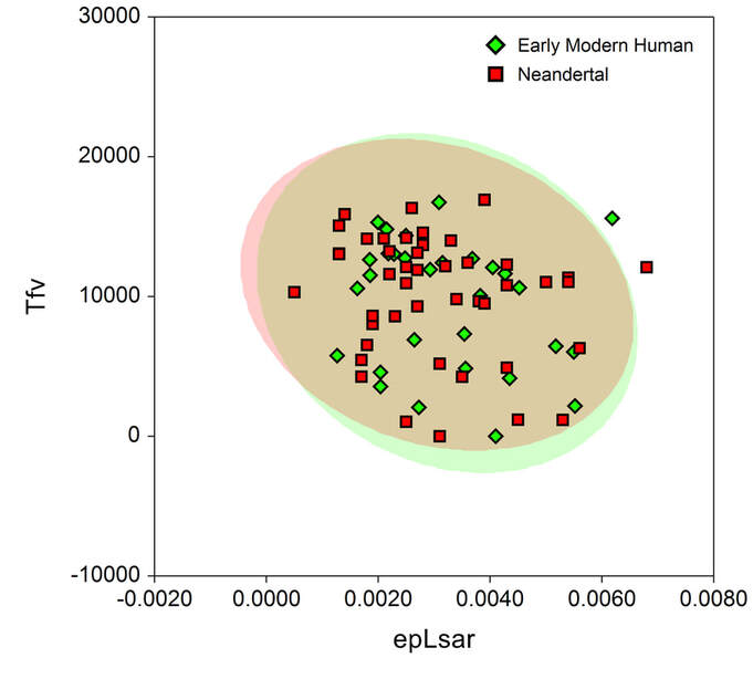 Above: Scatter plot of Neandertal and early modern human dental microwear texture values (Tfv = textural fill volume & epLsar = anisotropy) with 95% confidence interval ellipses. Note the extensive overlap in variation. 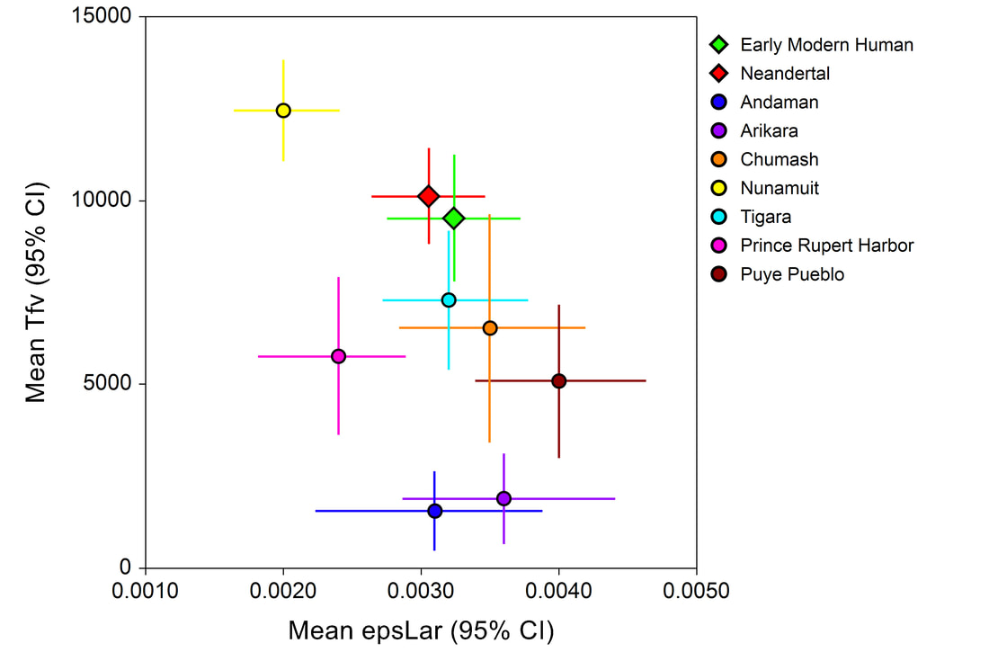 Above: Means and 95% confidence intervals for textural fill volume (Tfv) and anisotropy (epLsar) for the Neandertal, early modern human, and Holocene/ethnohistoric comparative groups. Note the closeness of the means Tigara group to the Pleistocene humans. The colloquial meaning of "neandertal" reflects the long-held notion of a major behavioral gap that separates the Neandertals from "us". However, the scientific and public perception of the Neandertals is undergoing a sort of renaissance at present. Many behaviors once associated with modern humans - like the controlled use of fire or creation of cave art and personal ornaments - are now documented in Neandertal contexts. The dental evidence presented here is also significant given the long-held view that Neandertals relied so heavily on their bodies, teeth, and brawn over technological and cultural solutions to complete their daily activities whereas early modern humans were thought to use brains, culture, and technology over brawn. However, the dental evidence tells a different story - there's little noticeable difference between the Neandertals and early modern humans in terms of tooth-using behaviors. I am of the opinion that the presumption of a major behavioral gap has persisted for so long because early modern humans were so infrequently compared directly to Neandertals in so many analyses. Instead, Holocene and/or ethnohistoric peoples were generally used (and sometimes still are) as proxies for early modern humans from the Pleistocene. While it should be obvious that historical and contemporary hunter-gatherers from disparate locations (common examples come from East and South Africa, the Arctic, and Australia) are not equivalent to Ice Age hunter-gatherers from Europe, uncritical comparisons were (and occasionally still are) the norm in paleoanthropological research. The work presented here shows how direct comparisons between Neandertals and early modern humans blur the divisions once thought to exist for anterior tooth-using behaviors. Whereas the data from recent human groups can be used to refine interpretations of Pleistocene behavior rather than be used as proxies for the early modern human behavior. Lastly, this publication is available freely (open access) and all data is freely downloadable in the online supplemental information. This was a really fun piece to contribute to and I am really proud of how it turned out. Many thanks to the first author for the chance to collaborate! Really looking forward to seeing what research Dr. Krueger sinks her teeth into next.... References FREE FREE FREE DOWNLOAD!
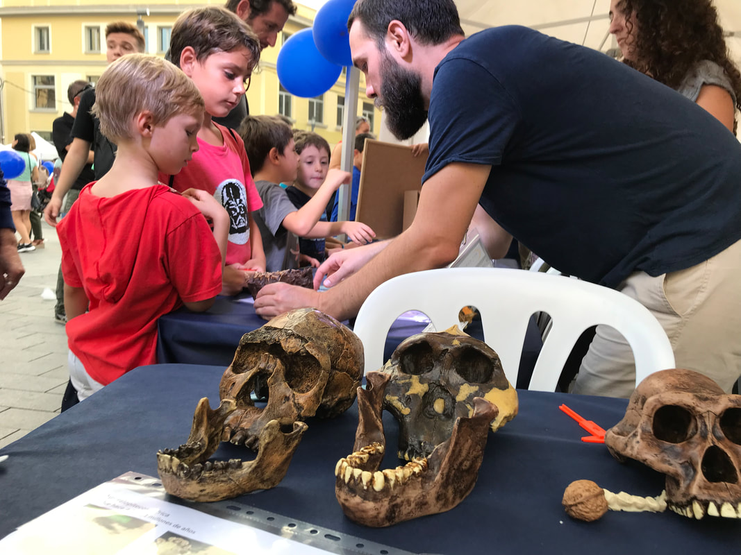
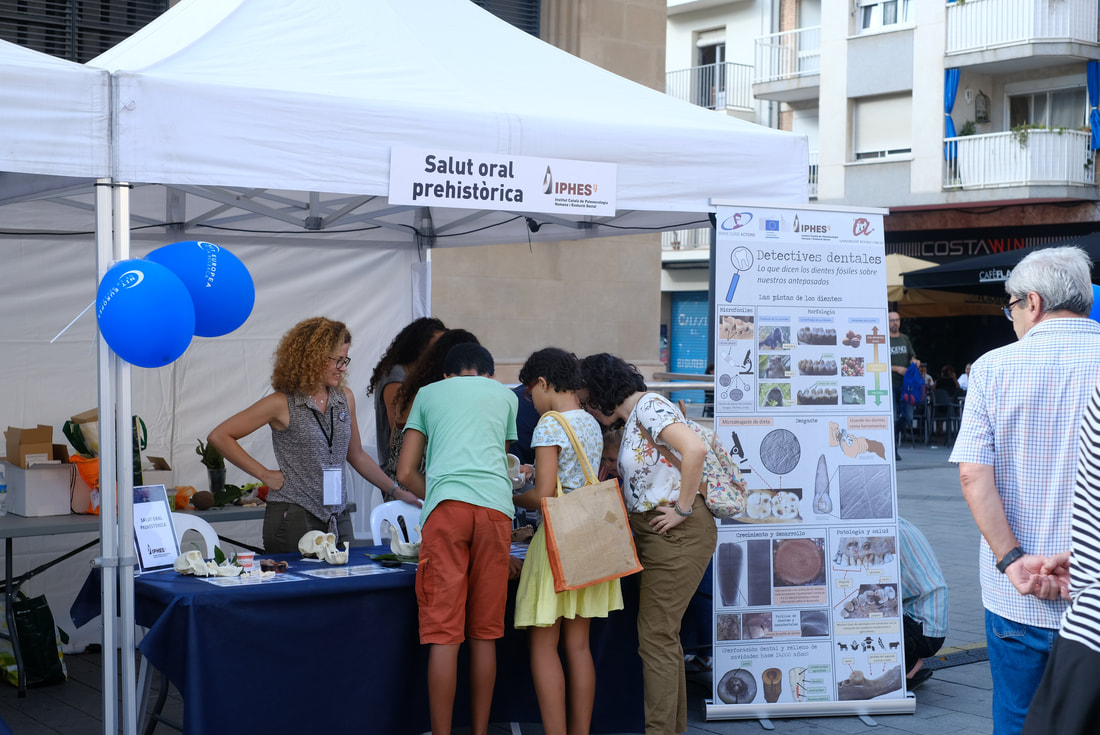
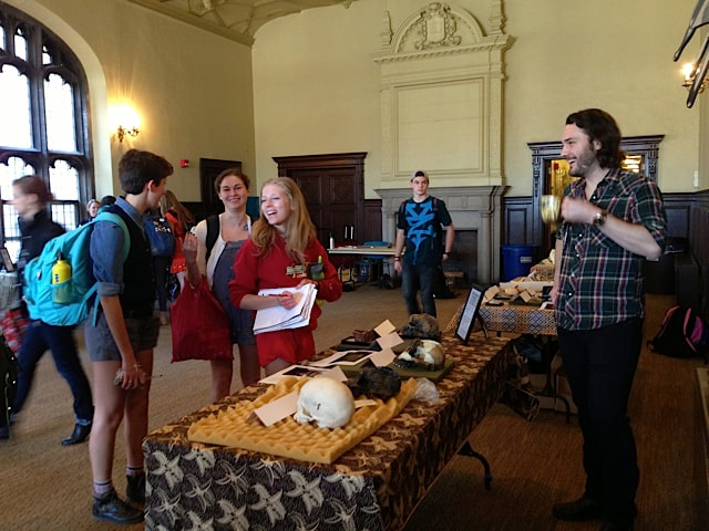
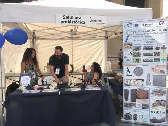
Krueger KL, Willman JC, Matthews GJ, Hublin J-J, and Pérez-Pérez A. 2019. Anterior tooth-use behaviors among early modern humans and Neandertals. PLOS ONE 14(11):e0224573, doi.org/10.1371/journal.pone.0224573. Krueger KL. 2015. Reconstructing diet and behavior in bioarchaeological groups using incisor microwear texture analysis. Journal of Archaeological Science: Reports 1:29-37, http://dx.doi.org/10.1016/j.jasrep.2014.10.002. Krueger KL, and Ungar PS. 2009. Incisor microwear textures of five bioarcheological groups. International Journal of Osteoarchaeology 20(5):549-560, dx.doi.org/10.1002/oa.1093. Krueger KL, Ungar PS, Guatelli-Steinberg D, Hublin J-J, Pérez-Pérez A, Trinkaus E, and Willman JC. 2017. Anterior dental microwear textures show habitat-driven variability in Neandertal behavior. Journal of Human Evolution 105:13-23, http://dx.doi.org/10.1016/j.jhevol.2017.01.004. A little over two weeks ago was the Nit Europea de la Recerca (European Researcher’s Night) in Tarragona. It was part of a massive initiative that involved events in over 300 cities across Europe. The theme centered on International Year of the Periodic Table. Several of my IPHES colleagues and I (Institut Català de Paleoecologia Humana i Evolució Social, IPHES) participated in the event alongside numerous researchers from the Universitat Rovira i Virgili (URV), Catalan Institute of Classical Archaeology (Institut Català d’Arqueologia Clàssica, ICAC), the Pere Virgili Institute of Health Research (Institut d'Investigació Sanitària Pere Virgili, IISPV), and Institute of Chemical Research of Catalonia (Institut Català d'Investigació Química, ICIQ) provided exciting and unique scientific outreach activities for the children and families. The event was free and took place in front of the central market (Plaça Corsini) in Tarragona. Tables and booths were set up for all the research groups and the children started pouring in around 4:30.  Our contribution was Detectives Dentales: Lo que dicen los dientes fósiles sobre nuestros antepasados (Dental Detectives: What fossil teeth say about our ancestors). Like the rest of the researchers, we had a booth with tables set up, hands-on activities for the kids, and a giant poster describing our research. Hundreds of children came to our table to learn about human evolution, primates, hominins, and bioarchaeology over the course of the night. Thanks to the support and help of my incredible colleagues (Marina Lozano, Raquel Hernando, Efstathia Robakis, Miquel Guardiola Fígols, and Marta Fontanals Torroja), and the organizers of the event, everything went incredibly smoothly. ComCiència URV (@cienciaURV) also provided some great pictures of the event: Admittedly, I was terrified of doing this event. My Castellano is terrible, and my Catalan is non-existent, which is a constant source of stress and embarrassment for me. While my colleagues made sure there were no horrible mistakes in the materials we used (poster, fact sheets, etc.), there was still the issue of needing to speak Castellano for about 5 hours. Thankfully, I had incredible help with the event from my colleagues. And then there were the parents and the children… Simply put, they were incredible! I spoke in Castellano (as best I could) for most of the event, but many parents would extrapolate what I was communicating to their children or translate it into Catalan. In fact, there was a lot of translation. Examples I can immediately recall included translating my Castellano to Chinese and Italian. Other children that were not originally from Spain, were more comfortable with English than Castellano, so I obliged. Some of these children even translated what I said (in English) to their mother tongue for their younger siblings – examples included German, Italian, and Chinese. Some children would just look at me puzzled when I spoke, then took a look at my name tag (misspelled “John Williams” for the night, but still very much not a Spanish name), which prompted them to ask, “Can you say something in English?” I wasn’t prepared for this, but it was quite funny, and gave some children a chance to practice English. Another common end to an interaction was a “thank-you” in English from parents as they moved on to another table – a kind of wink and nod that they appreciated the effort. All in all, was surprised at how well everything went despite my initial anxieties concerning language barriers. Some reflections on this type of public outreach event I wanted to share a few reflections on what worked well for this type of outreach event, since many of my colleagues are involved in these sorts of activities. I envisioned setting this event up much like the “fossil laboratory” events that I used to set up for Introduction to Human Evolution / Biological Anthropology courses when I was a graduate student – albeit, this was for a much younger audience! The target age group was around 5-12 year olds but we definitely had some younger children too. And to be honest, many of the parents showed an interest in the materials on par with, and sometimes exceeding, that of their children! So here’s what worked and didn’t:
An enormous poster: We had a giant poster with lots of examples, or “clues”, that biological anthropologists use to understand and interpret primate and hominin dental morphology, diet, growth and development, behavior, pathology, etc. By “giant”, I mean 200 x 80 cm… About three times larger than any conference poster I’ve ever made… It was a monster. Keep in mind that Powerpoint will not make posters this big, so a half-size (100 x 40 cm) poster was made. I had to constantly remind myself to zoom in 200% to get an accurate sense of whether or not each image would be blurry or if the text was too small. In the end, the poster turned out really well and will be used for future science communication events at IPHES since it was printed on durable vinyl.
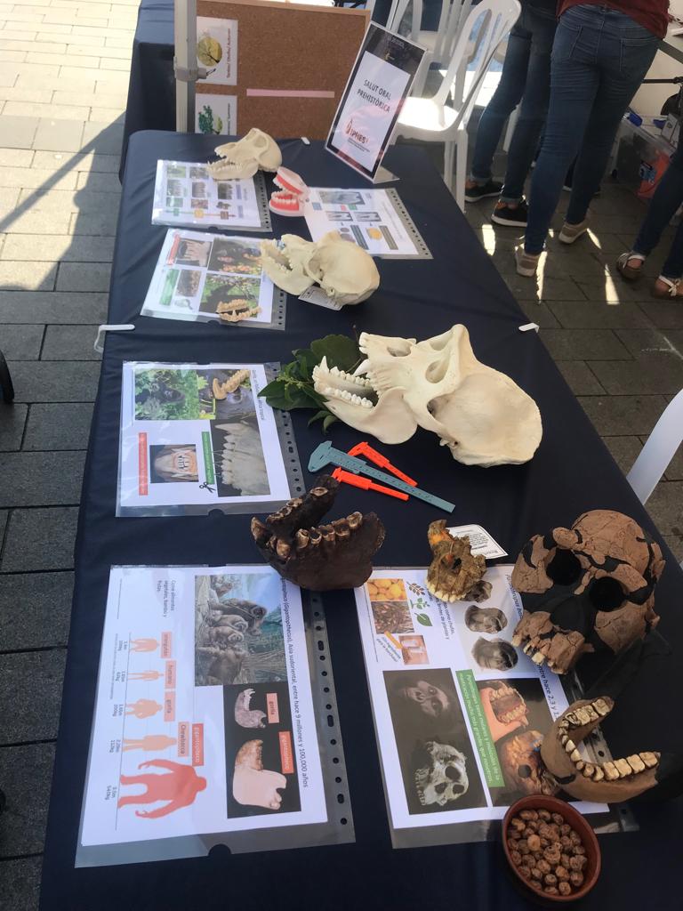
Primate and hominin casts (“No these are not the original fossils”): We set up a variety of casts from the URV/IPHES teaching collections to illustrate various aspects of primate functional morphology and hominin cranial and dental evolution. What I didn’t know would turn out to be so useful were the laminated sheets that accompanied each cast or groups of casts. For example, sheets for living primates included bright and colorful images of living primates eating and some basic descriptions of how their teeth help break down certain types of food. The hominin casts that we used included examples of Australopithecines, Paranthropus, Homo erectus, archaic Homo – a Neandertal and the Sima de los Huesos Skull 5 (“Miguelón”), and a recent modern human. Every cast also had an information sheet with a few facts about the fossil and reconstructions to help the kids visualize what these hominins may have looked like in life (The Smithsonian Institution, as always, was a great resource for this). The kids really liked the Gigantopithecus blacki mandible, but this was partly due to the comic relief provided by the body size comparisons in the information sheet we provided. The laminated sheets were not taped down, which also helped a lot. We were often completely surrounded by kids and unable to move around the tables with ease. Instead, being able to reach and grab the facts sheets and casts from across the table proved very helpful.
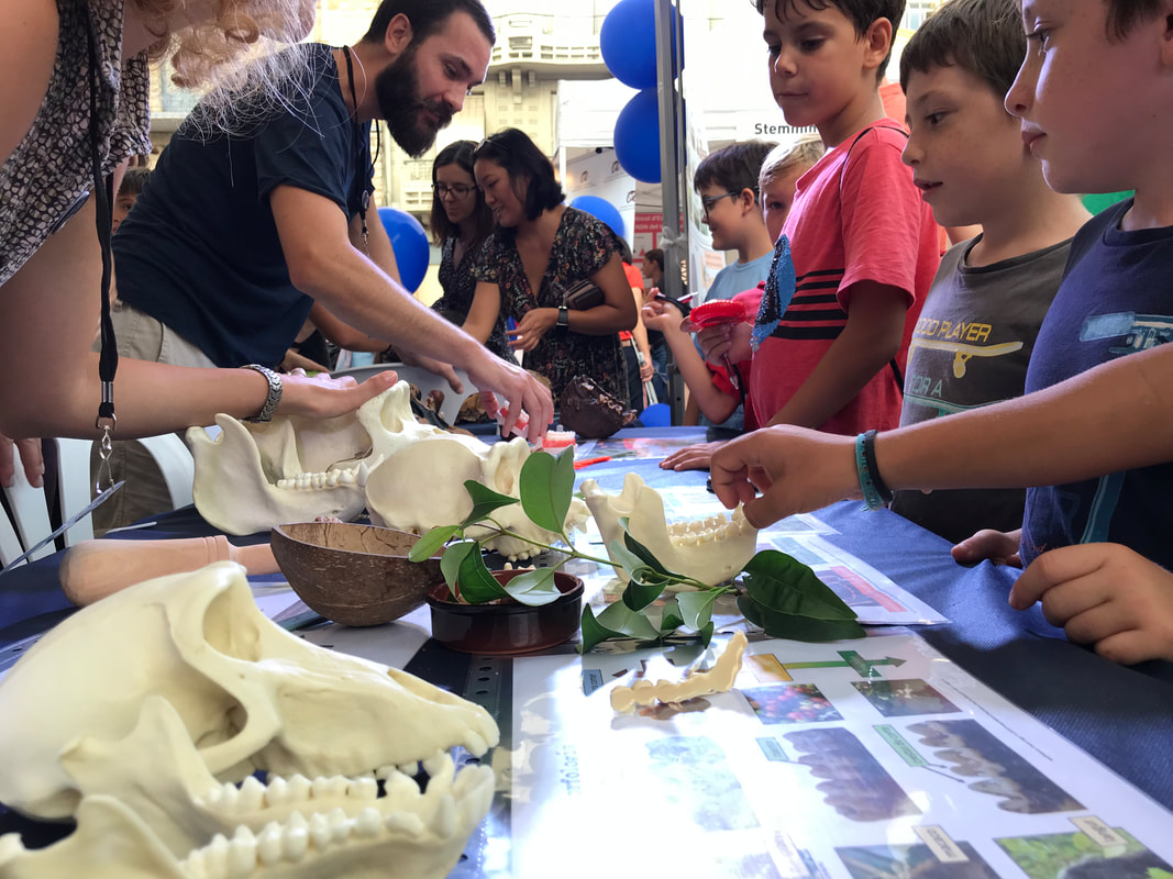
Food breakdown: To help demonstrate some of the ways that teeth breakdown food, we had some props on the table to help with explanations. We used dried figs as our fruit example since they are a much less messy alternative to fresh fruit. We paired the fruit with a mortar and pestle to discuss bunodont molars. We had plenty of leaves on the table, and we discussed shearing crests by miming the cutting action of scissors with our fingers. Originally, I was going to have real scissors to cut leaves but quickly realized how dangerous that might be as children started swarming the table! We also had walnuts (hard food), a nutcracker, and chufa (tough, high fiber food) on display. The chufa was one of my personal favorites and it was easily identified by parents (and some kids) as an ingredient for horchata. Aside from being delicious, chufa is a hypothetical C4 plant food of Paranthropus bosei – original article here). My colleague, Dr. Erin Kane, uses similar strategies in her course on the “Evolution of the Human Diet” which inspired me to go ahead with this idea at Researcher’s Night. She used a variety of other tools and foods for her demonstrations - like a staple remover to demonstrate how some teeth are well-suited for piercing the exoskeletons of insects - which are great for classroom settings. Like the scissors, we couldn't incorporate all the examples that Dr. Kane uses, but I can't wait to expand this activity the next time I get to teach this section in my own course. Dr. Kane also explores oral processing of food in her class with a variety of snacks – another thing I can’t wait to adapt for outreach events and teaching in the future.
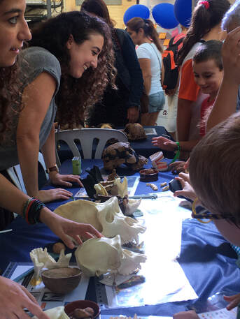
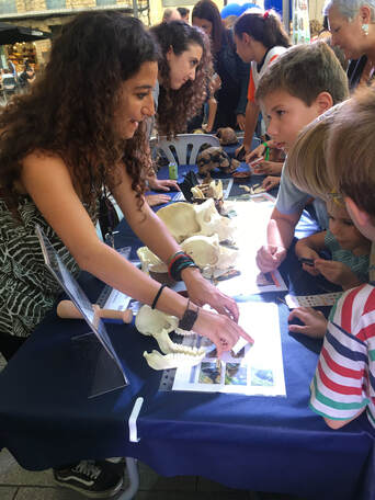
My colleagues, Raquel Hernando and Efstathia Robakis, teaching children about non-human primates and early hominins. If you look closely you will find a child playing with the loop. Other odds n’ ends: I molded the teeth of each fossil cast that we had on display and made dental casts in advance of the event out of a cheap and durable polyurethane resin. These casts were a lot easier for smaller children to handle. I also put a few 10x magnifying loops on the table and a handful of small plastic calipers. I never would have thought they’d be so popular, but many children measured everything they could – from the teeth and jaws on the table to the smile of a friend. The loops were even more popular and worked perfectly for examining the extra dental casts I made. One young scientist, I’m guessing she was 4 years old, came back to the table at least 3 times over the course of the evening just to use the calipers and loops! I rarely left my home without a loop when I was that young, and it is nice to see that these cheap little devices still fascinate children in an age of touchscreens.
What I would do differently: One of my big regrets centered around not having souvenirs for the children. This was largely due to a lack of time on my part and it’s something I definitely want to be better prepared for in the future. The original idea was to create a large sample of hemi-mandibles or hemi-maxillas out of dental stone. A variety of non-human primate and hominin teeth would be made, and each cast would come in a small bag with a fact card describing morphology, diet, etc. It’s not much, but I remember loving these sorts of things as a kid, and I would have liked to have had them ready for the kids at Researcher's Night. Alas, several hundred dental casts required quite a lot more time than I had to spare prior to Researcher’s Night. More loops. I ordered a 10 pack of loops but only one unit arrived, and I only had one other I was willing to risk damage to. The kids really liked them, so next time I will be better prepared with a dozen or so cheap loops spread across the tables. I’m sure I’ll continue to think of other things I’d wish to change but I think I’ll save that for the aftermath of the next public outreach event I take part in. All in all, this was one of the most unique and rewarding events during my postdoctoral experience in Tarragona. Again, this is in large part to the incredible help provided by my colleagues and the enthusiasm of the children and families that attended Researcher's Night. Many thanks to all! |
John C. Willman
A place to find updates about my research. Archives
July 2021
Categories
All
|

 RSS Feed

_-_Jos%C3%A9_Malhoa.png)